Sunday, January 22, 2012

Southern California Research Trip, Part 2: Natural History Museum of Los Angeles County (exhibits)

After two and a half days at the San Diego MNH, Morgan, Sarah, and I said our goodbyes to our friends and colleagues there (Joe El Adli, Eric Ekdale, and Tom Demere), and piled into my tiny honda for the drive up to LA - we decided that spending another morning at SDNHM would allow us to drive up to LACM and miss all the morning traffic. We arrived at the museum at about 2 in the afternoon, and after chatting with Curator Emeritus Dr. Lawrence ('Larry') Barnes for a little bit, we got right to work examining fossil pinnipeds. I'll discuss the collections visit in the next post - first I'd like to talk a bit about the new mammal paleontology hall.

The new "Age of Mammals" hall has been in the works for several years, and has taken quite a bit of time on behalf of most of the Paleontology Dept. employees at LACM. I've seen a few photos on the internet prior to my visit, so I knew a little of what to expect. I don't have much of a research interest concerning terrestrial mammals - so, sorry terrestrial paleomammalogists who happen to be reading this blog (admittedly a very, very, very small fraction of humanity), but I'm going to ignore the land mammals. Some of the marine mammal highlights include a mounted skeleton of the sperm whale Aulophyseter morricei from the Sharktooth Hill Bonebed, as well as the holotype skeleton of the phocoid pinniped Allodesmus kelloggi (which, according to some, may be a junior synonym of Allodesmus kernensis), and adult and juvenile mounted skeletons of the late Miocene dugongid Dusisiren jordani, which were collected from the Santa Margarita Sandstone in Santa Cruz County. Lastly, and arguably the centerpiece of the marine mammal exhibits - is a beautiful new skeleton of Paleoparadoxia (which apparently may be named as a new genus in the near or distant future). Aside from these, there are a handful of skulls and pinniped fossils on display, including the world's oldest delphinid dolphin fossil - a complete skull from the Monterey Formation, unnamed and still undescribed. Overall, however - I must admit I was a bit underwhelmed. Certainly on the lower floor, there are plenty of fossil land mammals just packed in. But the top floor, which was an odd mix of La Brea specimens and marine mammals, there was just a lot of empty space, and there wasn't really that much marine mammal material on display, which is surprising given that the LACM holds one of the largest fossil marine mammal collections in North America.

Not in the Age of Mammals hall, but I had to include a picture of my favorite
pinniped, Callorhinus ursinus.

The juvenile skeleton of Aulophyseter morricei from the Sharktooth Hill Bonebed. This individual is under 50% adult size. The only known skeleton of this taxon.

This also was not taken in the Age of Mammals hall - but it goes to show that my wife likes to photobomb fossils all the time. I mostly put this up here to demonstrate to others that she is an awful person.

The holotype skeleton of Allodesmus kelloggi.

A cast of the holotype (and only known specimen) skull and jaw of the desmatophocid pinniped Atopotarus courseni. Atopotarus has occasionally been recombined as Allodesmus courseni, but desmatophocid taxonomy will have to be covered in a separate post.

A referred lower jaw of the Miocene sirenian Dioplotherium allisoni.

The adult female and juvenile skeletons of Dusisiren jordani from the Santa Margarita Sandstone in Santa Cruz County. The juvenile is so damn cute...

A Paleoparadoxia ulna with sharktooth bite marks. This specimen belongs to the mounted skeleton shown below.

A rather bizarrely portrayed fossil dolphin in some artwork related to sharktooth bite marks; I'm sure the artist was more interested and familiar with depicting sharks than cetaceans.

One of the exhibits I was looking forward to as a taphonomist - the gut contents of a Basilosaurus cetoides skeleton from Mississippi! It had as gut contents when it died
a mass of fish bones.


The articulated forelimb of the late Miocene delphinoid Albireo whistleri, originally described by Larry Barnes from the Almejas Formation at Cedros Island in Baja California.

The business end of the new Paleoparadoxia skeleton.

I'll leave you with this large size image of the Paleoparadoxia skeleton - my dslr camera doesn't have a wide angle lens, so I had to stitch these photos together for an ultra-size photo (I have a much larger version; contact me if you want it). And yes, that is the intrepid Morgan Churchill standing behind its ass.

Wednesday, January 18, 2012

Southern California Research Trip, Part 1: San Diego Museum of Natural History

After a ten day visit with my wife's family for Christmas in Billings, Montana, my wife and I flew back to California, and had two days to get ready for a research trip to southern California with our friend and colleague Morgan Churchill. Except that we both got sick with a nasty two day cold - we got back on a thursday night around midnight, and were supposed to leave on sunday morning (which also happened to be January 1st). Needless to say, neither of us really were able to get ready for the trip on account of how crappy we both felt, and miraculously, waking up on sunday morning for the trip, I finally felt okay to drive, and the trip was a go (only we still had to pack, and didn't get out of town until close to 1pm). It's an 8 hour drive from San Francisco to San Diego (without traffic), so we were looking at a pretty late arrival. That being said, we had just driven up to Montana a couple weeks prior, and 8 hours seemed miniscule compared to two 10 hour days across the American west. We got to Los Angeles around 7pm, and stopped in for a couple hours to visit our best friend, Lee Hall, and his awesome girlfriend Ashley Fragomeni, which was refreshing (and far too brief). We finally pulled into east San Diego to pick up Morgan at about 12:30am (after driving through the densest coastal fog I've ever seen - and that's coming from a San Francisco native!). We pulled up to the house of our gracious host (and my coauthor) Joe El Adli, who also hosted me during the SATLW meeting in June at SDSU.
Morgan measuring a skull of Valenictus chulavistensis, the "toothless" walrus from the Pliocene San Diego Formation.

We had several goals for our visit - Morgan and I are studying a new specimen of the middle Miocene walrus Pelagiarctos, which Larry Barnes descibed in 1988 from the "chin" end of a pair of mandibles and some teeth, and suggested some interesting hypotheses regarding its paleoecology. At San Diego in particular, Morgan needed to spend some time photographing and measuring every pinniped skull and jaw he could (and it took up nearly the entire time of our visit). When not helping Morgan with measurements, I was chatting with Tom Demere (paleontology curator at the SDNHM) and Joe El Adli (lead preparator of the paleo department at SDNHM) about the fossil mysticete Herpetocetus, as well as photographing some earbones and crania of balaenopterids that occur in both the Purisima and San Diego Formations.

A mounted skeleton of Allodesmus (kernensis? gracilis? depends on who you talk to) at the SDNHM, while the tail and claspers of a giant Carcharocles megalodon loom ominously above.

A closeup of the business end of Allodesmus.


A skull of Desmatophoca oregonensis, a smaller and earlier relative of Allodesmus from the Astoria Formation of coastal Oregon. This specimen was collected by Douglas Emlong, and described by Tom Demere and Annalisa Berta in 2002.

Two pinniped skulls in particular that we definitely needed to see are on display in a huge cabinet out in the Fossil Mysteries hall. During my 2007 visit, I had been let into the cetacean display case to examine and photograph a porpoise skull, but that case actually had an entire door and you could walk around in it; this display case with the walruses had a 150 pound sheet of glass that required four of us (Morgan, Joe, myself, and the SDNHM collections manager, Kesler Randall) to use large suction cups to lift the ~7 foot tall sheet off of the case (which required Joe and Kesler to go up on ladders on either side). It was quite the performance, all done prior to the museum opening.

Joe (left) and Kesler (right) brainstorming about removing the sheet of glass from the walrus exhibit.

Joe placing the suction cups on the glass. The two walrus skulls in
question
- Dusignathus seftoni on the left, and Valenictus chulavistensis on the
right - can be
seen at the bottom of the case.

We needed to see two specimens in particular - one is the nearly complete and well preserved paratype skull of Valenictus chulavistensis, and the other is a referred skull of the dusignathine "double tusked" walrus Dusignathus seftoni. I had really ought to blog about each of these taxa, as they are truly wonderful and bizarre creatures. The new skull of Dusignathus was collected about 6 or 7 years after Tom Demere described and named the species in 1994, and is in much better shape than the holotype specimen, which is substantially smaller, and is missing the palate (although this new larger male specimen is missing the top of the skull). Although I've seen this display several times before, I must admit I was very surpised with 1) how small the Valenictus paratype is in comparison to other skulls, 2) how light the paratype is (Tom stated that it was very poorly mineralized and held together primarily with consolidant), and 3) how damn huge the Dusignathus skull is in comparison to other skulls in collections.

Joe (in brown) and Kesler (left) set the walrus crania down onto foam, while Morgan (right) and I (gray) watch.

While Morgan was taking photos, I decided to get a different angle on some of the fossils while I had a ladder available. Here I am shooting the Allodesmus photos seen above.

Here's a nice shot I got from the ladder of a new species of balaenopterid mysticete from the Pliocene San Diego Formation (this taxon is being studied by SDSU student Jessica Martin). So far as I can tell, I've not yet seen this animal from the Purisima Formation.

A neat mount of a fossilized wing of an albatross, Diomedea sp. (but should probably be identified as Phoebastria sp.). Albatrosses are definitely huge birds - but the partial humerus of Pelagornis sp. I recently published, which was only 1/2 complete, was the same length as the complete humerus of this specimen.

My wife always makes friends on research trips.

By the end of the San Diego visit, we had each taken several gigabytes of photos, and dozens of measurements for our research. My wife had spent a fair amount of time next door at the San Diego Zoo, and at the Museum of Man. We spent wednesday morning at the SDNHM to wrap up everything before driving to LA, where we would spend our first afternoon of research at the Natural History Museum of Los Angeles County (NHMLAC), but known to paleontologists simply as "LACM" - which will be the subject of the next post.

Saturday, January 14, 2012

New USB microscope

As previously mentioned, I've been using a USB powered microscope recently, which I purchased on Amazon for about 60 bucks. The Veho Discovery VMS 004 deluxe is tiny (a little larger than a cigar), and plugs right in to a USB port - I've already used it at UC Berkeley on a couple visits to photograph tiny vertebrate bones and teeth for a colleague. I had about 70-100 specimens I needed to photograph, all under 1cm in length, and using the microscope which was plugged in to my laptop, I was able to take photographs of all the cardinal anatomical directions in about three hours (~300 photos, so a rate of a minute per photo). Unlike a digital camera, the microscope immediately shows you if the picture turned out or not on a relatively large screen (digital photography has sped up picture taking so much that I occasionally get careless, and unintentionally let one or two photos out of every hundred get blurry and crappy, which is less of an issue with a larger display screen).

At Nick Gardner's request, I've posted some examples of USB photos I've taken. It's not the highest quality in terms of photography, and sometimes some of the images appear a little pixelated. Most of the time, the photos look okay, if not on the smaller end of publishable quality (i.e. smaller in terms of file size and resolution). Furthermore - don't buy this product if you're expecting continuous zooming, which they advertise that it has, but it really doesn't. It really only has two focal points, which is a problem. But then again, this thing is only 60 bucks, so you get what you paid for. All in all for the money I spent, I'm fairly satisfied as it effectively acts as a low-price, portable electronic dissecting scope. Lastly - the photos I've uploaded have not been modified in any way, so you can get an idea of what to expect.

Edit: I forgot I should add this in here - on the Amazon page for this item, there is a pretty lengthy review of the product by Paul Clifford, who has graciously written his own impromptu user manual for the microscope. He discusses some of the finer technical points a bit more technically than I have, and I'll admit I found his review very helpful when trying to experiment using this thing.

Most of these photos would not be high-resolution enough to be published at full page or even column width size - *but* would probably work well in a situation where you're figuring a number of small specimens all on the same figure.

An oral tooth of a wolf eel, Anarrhichthys sp. from the late Miocene Wilson Grove
Formation of Sonoma County.


A lateral tooth of a bat ray, Myliobatis sp., also from the Wilson Grove Formation. Yes, that is play-doh that Pat Holroyd gave me to photograph on; they don't like using clay at UCMP because it has nasty oil in it.

The apical view of the same Myliobatis sp. tooth.


Lastly, for all you cetaceaphiles, here is the stapes of UCMP 219111, a well preserved cranium of Herpetocetus bramblei from the Purisima Formation of Northern CA.

Up next - tales from my recent southern California museum collection visit with my wife and Morgan Churchill, to examine fossil pinnipeds at the San Diego Natural History Museum and the Natural History Museum of Los Angeles County.

Saturday, January 7, 2012

Dead baby fur seals at the California Academy of Sciences

Well, it’s been over a month since I’ve posted anything. It’s been a busy winter – in the mean time, my wife and I have celebrated Thanksgiving in California with my family, and have been in Montana for the last week with her family. I’ve been typing like a beast (30 pages for one manuscript just since I’ve been back in Big Sky country) over the last month or so. I’ve been working on my longest manuscript yet (not including my thesis, however it will soon overtake it), which as of this morning reached 110 pages. Additionally, we’ve been trying to get certain things ready for our imminent move to New Zealand. Just prior to Christmas vacation, I spent a couple of days visiting the Ornithology and Mammalogy collections at California Academy of Sciences.

A menagerie of bird and mammal skeletons and mounts.

A giraffe skull sits next to the sign in book.

A row of shelving filled with fur seal and sea lion skulls and skeletons.

A beautiful mounted skeleton of a sea otter (Enhydra lutris).

I’ve been making a couple visits per year to Cal Academy since 2006; I originally visited when the new building was under construction, and the academy (exhibits, departments, and all) were at the temporary storage facility in SoMa (South of Market in Downtown San Francisco, for the non Bay Areans) to check out their collection of Purisima Formation fossils, and to utilize their ichthyology and mammalogy collections to identify shark teeth and pinniped bones from various Miocene and Pliocene strata from Northern California.

A bunch of large mysticete vertebrae awaiting curation.

A pilot whale (Globicephala macrorhinchus) skull in the CAS collections.

On previous visits, I’ve searched the Mammalogy collection of skeletons to make comparisons with modern and fossil bones and teeth of fur seals, walruses, pilot whales, porpoises, and baleen whales. In 2010, I collected some (relatively basic) data on the variation of tooth root lobe morphology in northern fur seals (Callorhinus ursinus) for comparison with fossil fur seals, which I published in JVP earlier this year. A current project I am working on is writing up an entire marine vertebrate assemblage (~200-300 fossils), and I am working on a lengthy manuscript on the marine mammal compliment of the assemblage. Needless to say, the large and very well curated collection of marine mammal skeletons at California Academy of Sciences has been indispensable throughout this endeavor, and has made many fossil identifications possible and paved the way towards insights into marine mammal osteology.

Many crania and jaws of the Northern Elephant Seal (Mirounga angustirostris) are too large for storage boxes and sit right on the shelves.

A walrus (Odobenus rosmarus) skull with two baculi thrown in for good measure (no pun intended).

A bunch of boxes full of Galapagos Sea Lion skeletons (Zalophus californianus wollebaeki).

On my most recent visits, my objectives were threefold: first, to take photographs of many extant species of otariids (fur seals and sea lions) for a morphobank project with my colleague Morgan Churchill; second, to photograph nearly every skeletal element from an adult northern fur seal in order for comparison with fossil fur seals from California (e.g. Thalassoleon, which has previously been hypothesized to be closely related to Callorhinus); and third, to photograph and examine lower jaws of neonatal and fetal fur seals and sea lions.

Several drawers filled to the brim with boxes of small (fetal, neonatal, and juvenile) otariid skulls. Most of these are Callorhinus ursinus.

I won’t get into the specifics quite yet, nor will I talk about the fossils that spurred my curiosity regarding the third subject – I’ll only say that it is pretty damn neat if I may say so myself. That being said – I am very interested in the morphology of deciduous (milk) teeth in young fur seals, as well as the timing of molar and premolar eruption in the lower jaws of these animals. I’ll briefly mention that modern pinnipeds are a bit weird in that they (like most) mammals have milk teeth, but they are often shed before birth, so that the pups are born with a full set of adult chompers. Their milk teeth have a very reduced functional period, and additionally are reduced to tiny little pegs (unlike the milk teeth of terrestrial carnivores). Although they still develop milk teeth, pinnipeds are trending toward monophyodonty – that is, having only one set of teeth as opposed to two (diphyodonty). Cetaceans are monophyodont, and pinnipeds are an excellent example of a second clade of marine mammals following the same evolutionary trend.

A baby fur seal head (Callorhinus ursinus).

A neonatal Callorhinus ursinus skull in lateral view.

In order to examine the tiny milk and permanent teeth of these pups and fetuses, I brought along my new toy – a small, portable, USB powered digital microscope which plugs into my notebook laptop (…another new toy, which I’m using from a secure location in Montana). It displays the image on the screen, and can acts as a camera as well. Fortunately, there is a button to take a picture with in the software, rather than having to manually press a button on the microscope (which, due to its small size, usually jiggles it and screws up the picture). At an earlier UCMP visit in October, I was able to take around 200 photos of 100 tiny fossil specimens in a little over two hours. With the digital microscope, I was able to take a bunch of photos of milk and permanent teeth from nearly a dozen or so specimens of northern fur seal (Callorhinus), California sea lion (Zalophus), and Steller’s sea lion (Eumetopias). Unfortunately, there weren’t any northern fur seal fetuses, or specimens with deciduous premolars – but the data for sea lion fetuses I collected was more than sufficient to answer my fossil-related queries.

Yours truly using my digital USB microscope to examine the morphology of
fetal and neonate fur seal teeth; this specimen is a Northern Fur Seal (Callorhinus ursinus).

Milk teeth and unerupted adult teeth in the lower jaw of a California sea lion (Zalophus californianus) fetus.

Monday, January 2, 2012

Is Puijila a pinniped?

Back in February 2009, Natalia Rybczynski and colleagues surprised the paleomammalogy community with their Nature paper naming a new genus and species of early pinniped, Puijila darwini. The holotype skeleton is relatively complete, and include fore- and hind-limbs along with much of the vertebral column, both jaws, and a well preserved skull. Puijila was about one meter long, and would have appeared relatively similar to a modern river otter. It had a short snout and a wide head, with large eyes and relatively high-crowned teeth. The teeth of Puijila still retain many of the cusps lost in modern pinnipeds, and also exhibit pits in the roof of the mouth for the lower teeth (embrasure pits). Unlike modern pinnipeds, it had a long tail, and did not have its fore- and hindlimbs modified into flippers. Puijila was discovered in 2007 from the Haughton Formation on Devon Island in Nunavut (formerly Northwest Territories in Canada). The Haughton Formation was deposited in an impact crater – the impact has been dated to 24-21 Ma (earliest Miocene), and fossil mammals from the Haughton Formation corroborate an early Miocene age. The Haughton Formation was deposited in an ancient lake that filled in the impact crater. In fact – if it were not for the impact, there would be no sedimentary rocks of Miocene age preserved that far North – all the young rocks have been eroded away by glaciation.
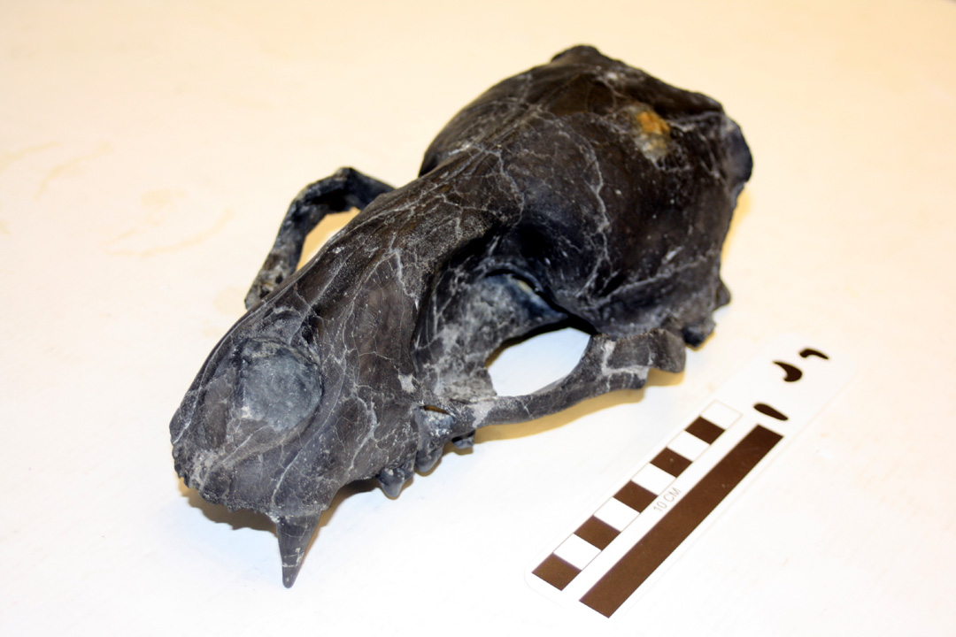
The skull and jaw of Puijila darwini, from Rybczynski et al. (2009).

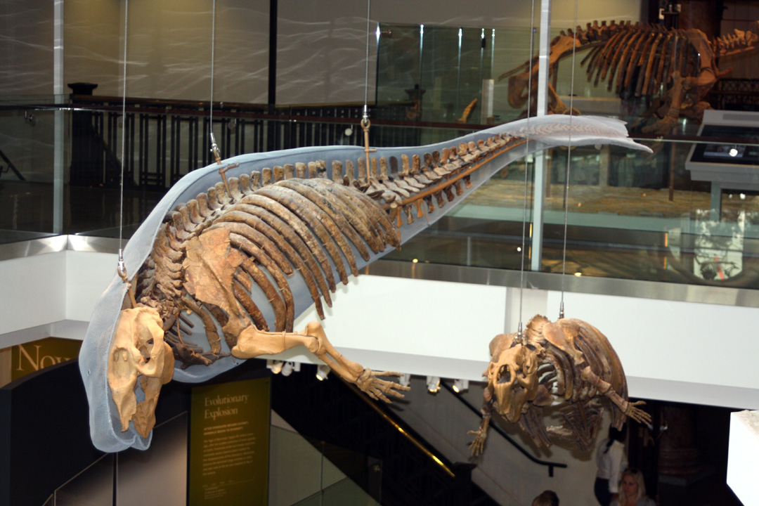
The holotype skeleton of Puijila darwini (from http://nature.ca)

Previously, the earliest diverging pinniped (and arguably more derived than Puijila) is Enaliarctos, a fur seal sized pinniped from the latest Oligocene and early Miocene of California and Oregon. Enaliarctos retains carnassials, although many of the other dental features are very simplified and reduced, trending toward the condition in modern pinnipeds. Enaliarctos also exhibits limbs developed into flippers – and is very clearly a pinniped. But the relationships of Puijila – to pinnipeds and other carnivores – are not so clear. Because of the older age of Enaliarctos and its marine occurrence, Puijila is hypothesized to represent a lineage of early pinnipeds that stayed in their freshwater niche while marine pinnipeds like Enaliarctos evolved, remaining otterlike. It suggests that pinnipeds went through an otterlike freshwater stage before invading the ocean. Prior to this, Enaliarctos suggested a direct to saltwater invasion – although due to the absence of intermediates, it was not exactly clear one way or the other.

The skeleton and life restoration of Enaliarctos mealsi, from Berta et al. (1989).

Before we continue – I must also be specific about some clade names. Although Rybczynski et al. (2009) refer to Puijila as a member of the Pinnipedia – which is not really the traditional cladistic nomenclature for basal pinnipeds. Annalisa Berta and colleagues (1989) proposed the clade Pinnipedimorpha, for Enaliarctos and all later diverging pinnipeds. Berta (1994) later proposed the name Pinnipediformes for Pteronarctos and all later diverging pinnipeds. Pinnipedia is nested within Pinnipediformes, and Pinnipediformes within the Pinnipedimorpha. Within this traditionally accepted and utilized framework, Puijila’s obviously more primitive morphology than Enaliarctos indicates it should be referred to as a pinnipedimorph.

Rybczynski et al. (2009) listed six characteristics that unite Puijila with Enaliarctos and other pinnipeds. These are: a posteriorly expanded palate (the palate extends posteriorly past the upper toothrow in pinnipeds), an enlarged infraorbital foramen (occurring within pinnipeds due to larger whiskers and greater innervation of the snout), a shelf-like protocone on the upper fourth premolar (occurring in basal pinnipeds and some related arctoids), an upper second molar that is reduced and positioned medially to the upper first molar (reduction of the molariform teeth to conical teeth is a major dental transition within the pinnipedimorpha), a posterodorsally expanded scapula (a feature of pinnipeds, which often have very broad scapulae, an adaptation for swimming), and an expanded deltopectoral crest of the humerus (another feature in pinnipeds related to swimming).

Some of these features may be of only limited use in hypothesizing a close relationship between Puijila and pinnipeds. First, an enlarged infraorbital foramen (the small hole below the eye socket in a skull) also occurs in many mustelids, such as badgers (Taxidea), weasels (Mustela), and most (if not all) otters (e.g. Lontra, Enhydra); in badgers and weasels, this is due to a more innervated and sensitive snout, an adaptation for digging in burrows. This characteristic may not be useful in identifying early pinniped relatives, as otters (another hypothesized pinniped sister taxon) exhibit this feature – presumably evolving for the same purpose. The two postcranial features – an enlarged deltopectoral crest and a posterodorsally expanded scapula – may not be coded correctly in Puijila. To be quite honest, based on the available photographs – these features do not appear too different between Puijila and a river otter (Lontra), and don’t really exhibit the derived condition, which is much more extremely developed in Enaliarctos and other fossil and modern pinnipeds). This leaves only three reliable characters behind.

Skeletons of Enaliarctos (A), Puijila (C), and Lontra (D).

One of these features – a posteriorly expanded hard palate – also appears in the aberrant arctoid Kolponomos, as well as Pachycynodon and Allocyon (Tedford et al., 1994). This feature appears to be more widely distributed than previously admitted, although it is worth mentioning that it is probably an adaptation towards aquatic feeding. The two dental characteristics are probably more reliable, although it is odd to note that these two dental characteristics are also found in Kolponomos. Kolponomos will feature more in part two of this post, as it is both a fascinating creature which I have not yet properly covered on this blog, and it is also extremely topical to the subject of Puijila.

A wonderful reconstruction of Kolponomos by Ken Kirkland, published in Neptune's Ark by David Rains Wallance.

There are a whole host of pinniped characteristics that Puijila does not have, or features that were not described in enough detail to independently evaluate. Cranial and dental features that characterize pinnipeds (either at the level of the Pinnipedimorpha, Pinnipediformes, or Pinnipedia) include a reduced upper first molar, reduced cingulum on the upper first molar, an absent or reduced metaconid, loss of the embrasure pit between the upper fourth premolar and first molar, closely spaced mastoid and paroccipital processes, a reduced nasolabialis fossa, reduced cingulum on the upper third incisor, and a reduced trigonid cusp on the lower molars. None of these derived conditions appear to be present in Puijila. A number of postcranial features which characterize pinnipeds (either at the level of the Pinnipedimorpha, Pinnipediformes, or Pinnipedia, as above) include a short and robust humerus, enlarged tuberosities of the humerus, a flattened radius and ulna that are longer than the humerus, emphasis of certain digits in the hand and foot (i.e. largest digit in the hand being the first or “thumb”, and the lateral and medialmost digits of the foot being largest and the smallest being the middle digit), a short, flattened femur with an enlarged greater trochanter and medially inclined condyles, a reduced tail, and a short ilium (bone in the pelvis). Curiously, none of these features are present in Puijila – although nearly all of them (perhaps all – I can’t remember off the top of my head) occur in Enaliarctos.

In summary, there are very few features that actually unite Puijila and pinnipeds. The cladistic analysis of Rybczynski et al. (2009) was relatively limited, both in terms of the number of fossil and modern species used, and also with regard to the number of morphological characters used in the analysis. An earlier study – considered to be the landmark phylogenetic analysis of pinnipeds, Berta and Wyss (1994) – utilized 143 characters, while Rybczynski et al. (2009) only used 42. Granted, the earlier study focused on relationships within pinnipeds – and many of those characters may or may not apply to Puijila and other similar arctoids.

Because of the above, I am skeptical that Puijila has a close relationship with pinnipeds – and although I’m not convinced, I am equally receptive to the idea pending further analyses. There is much we’ve been waiting for – a detailed description of its anatomy and large, detailed figures of its skeleton were not included in the original study, which is understandable given the short length of Nature papers (however, it was not included as supplementary info either). There is certainly more work to be done, and there are rumors of a more detailed phylogenetic analysis in the works; such a study should include a comprehensive description of the skull and skeleton of Puijila accompanied by exhaustive figures (formerly difficult, but several modern journals have made this a far less difficult prospect), sample a broader variety of caniform carnivorans as well as more fossil and modern pinnipeds (fossil pinnipeds with postcranial skeletons such as Allodesmus, Thalassoleon, Leptophoca, Acrophoca, Piscophoca, Imagotaria, Neotherium, and Proneotherium) as well as the “beach bear” Kolponomos (which was not included in the original analysis or discussed by Rybczynski et al. 2009). Puijila certainly could belong to some group of otterlike “protopinnipeds” as hypothesized, but given the few characters uniting them, it very well could be some sort of mustelid or other aquatic carnivore.

Further reading

Official Puijila website (Canadian Museum of Nature)

Puijila, a very basal 'pinnipedimorph' (this blog)

Commentary by Ed Yong (Not exactly Rocket Science)

Commentary by Brian Switek (Laelaps)

Literature cited

Berta, A., C.E. Ray and A.R. Wyss. 1989. Skeleton of the oldest known pinniped, Enaliarctos mealsi. Science 244:60-62

Berta, A. 1991. New Enaliarctos* (Pinnipedimorpha) from the Miocene of Oregon and the role of "Enaliarctids" in Pinniped Phylogeny. Smithsonian Contributions to Paleobiology 69.

A. Berta. 1994. New specimens of the pinnipediform Pteronarctos from the Miocene of Oregon. Smithsonian Contributions to Paleobiology 78:1-30

Rybczynski, N., Dawson, M.R., Tedford, R.H. (2009). "A semi-aquatic Arctic mammalian carnivore from the Miocene epoch and origin of Pinnipedia". Nature 458 (7241): 1021–24

R. H. Tedford, L. G. Barnes, and C. E. Ray (1994). "The early Miocene littoral ursoid carnivoran Kolponomos: Systematics and mode of life". Proceedings of the San Diego Society of Natural History 29:11-32.

Tuesday, December 27, 2011

October pinniped excavations

Dick Hilton and I excavating a pair of fur seal (Thalassoleon) dentaries from the base of the cliff.

A famished yearling California Sea Lion on the beach.

The weekend before the annual meeting of the Society of Vertebrate Paleontology in Las Vegas, my wife and I joined Dick Hilton from Sierra College for what we assumed would be a routine visit to the Purisima Formation at Point Reyes. We drove up on a Friday night, and cooked hot dogs at the campground with Dick, and set up our tent for the weekend. The following morning, we set out along the shoreline, and within five minutes, found what appeared to be a pinniped skeleton. Since we wanted to get on to more prosperous localities further down, we left it there with the intention of digging it up the following day (Sunday).
My wife pointing to something tiny she is proud of finding...

It's a beautiful fur seal (Thalassoleon) molar! She has a real eye for finding things like tiny pinniped and shark teeth, which is why I keep her around.

Dick Hilton trying to explain something to my wife and I. At least I hope he was talking to my wife, because it sure looks like I wasn't paying attention.

As we headed further on down to the good section of shoreline, I found a well preserved porpoise earbone (petrosal), and a few other odds and ends. Not too long afterward, and nearly at the same time, Dick spotted a couple bones eroding out with an associated tooth, and my wife spotted a well preserved fur seal tooth (Thalassoleon), only a few meters away. It took us the better part of an hour to dig the bones up – which turned out to be a pair of associated fur seal dentaries! Unfortunately, neither specimen had any cheek teeth, but one did have a broken canine. Either way, only the middle chunk of the dentary is preserved in the holotype of Thalassoleon macnallyae (from the same locality), and these are the most complete and well preserved jaws of this taxon now known. We hiked down the beach a little further, and collected a couple of pinniped limb elements – a proximal end of a tibia, and a metatarsal. After collecting these, we headed back towards the cars. On our way back, I spotted a string of a dozen articulated pinniped vertebrae – a second skeleton we would have to excavate the following day.

A cast of the holotype skeleton of Parabalaenoptera baulinensis on display at the visitor center.

For comparison, there is also a skeleton of an adorably tiny (~15 feet long) minke whale (Balaenoptera acutorostrata) on display as well.

My wife examining some baleen (presumably from a balaenopterid) at the visitor center.

We also finally had a chance to visit the visitor’s center; previously, I had always been there on a weekday, when the center was closed. I was particularly excited to check it out because it has one of the only known casts of the holotype skeleton of Parabalaenoptera baulinensis (which was excavated from the Santa Cruz Mudstone at a nearby locality) on display – there is one other, but it is at the College of Marin in Kentfield, and is falling apart and badly needing repairs.

My wife decided she was done, and decided to spend the day reading A Game of Thrones on the beach and napping and taunting birds all day.

On the second day, we spent about seven hours excavating the first pinniped skeleton, which resulted in about 100 pounds of tin-foil jackets. This skeleton is probably of a large fur seal or small walrus, and the skeleton was completely disarticulated; as soon as we were close to removing one bone, another would be under it, or behind it. There were probably about two dozen or so bones in the cliff that we excavated. At about 4 in the afternoon, we wrapped up the first excavation, and walked down the beach to relocate the articulated skeleton. It took a while to relocate it, but as soon as we did, we started excavating it in a large block. Thanks to the specimen being articulated, we were able to finish this excavation in a little over an hour. Once we got back to the car, we loaded up nearly two hundred pounds of fossils we had collected in only two days in the field; furthermore, the most ridiculous aspect of the weekend was that by weight, cetacean fossils comprised less than 1% of our haul (only one specimen). For the uninitiated – pinniped fossils are relatively rare, and my master’s thesis sample of specimens indicates that there is a 4:1 ratio of cetacean fossils to pinnipeds, and that pinnipeds constitute only 8% of the marine vertebrate assemblage from the Purisima Formation at Santa Cruz. To collect a pair of associated dentaries, a tooth, a couple of associated hindlimb bones, and two skeletons (one being articulated) all in one weekend – is surprising, and tripled the number of major pinniped finds I’ve made. I’ll post some updates when some of this material gets prepared.

She nearly got this gull to come to her backpack. Sorry, no pictures of the pinniped excavation.