Showing posts with label odontoceti. Show all posts

Thursday, April 5, 2012

Visit to the Otago Museum

I know there are some readers of this blog who have patiently waited and waited for pictures of beautiful Oligocene marine mammal fossils - to you I say, sorry for the delay. I'm going to try and get several blog posts written this weekend so I can post them incrementally. This one will mostly be in 'slideshow' format.

I've been fairly busy since I got here, and I've bordered on stress trying to figure out 1) where all the eomysticetid specimens are in collections, 2) which earbones belong to which skull or skeleton (just taking a while to become familiarized with the specimen numbers), 3) trying to make some sense out of the earbones and trying to group them based on consistently seen characteristics (and I have made a bit of headway), and 4) just generally trying to figure out how many taxa I am dealing with and thus 5) how many manuscripts/dissertation chapters this will end up making. Since I've finally made some headway and started describing the first material (a partial skull with earbones and a very partial postcranial skeleton), I've relaxed a bit and can allocate time to other activities. That being said, I'm also locked out of the building for four days due to construction/maintenance activities in the building. Fortunately, this will give me an opportunity to divert some time to my Pelagiarctos study with Morgan Churchill. Also, in other news - I finally finished up my massive manuscript describing an entire marine mammal assemblage from a locality in the Purisima Formation, which resulted in being just over 200 double spaced pages long with 45 figures; Felix Marx graciously offered to take a look, as did Ewan Fordyce. I have a bit of work left cleaning up some figures, but it should be submittable soon.
A spectacularly beautiful dalpiazinid dolphin! Look at those damn teeth! There's another specimen with even crazier incisors, and a full dentition, and jaw.

An archaic edentulous mysticete which may fall somewhere on the cetacean family tree near eomysticetids. This specimen will be part of my dissertation.


The holotype skeleton of the giant moonfish Megalampris keyesi. This set of slabs is seriously about 15 feet long and about 8 feet wide. Described by Gottfried et al. 2006.

A disarticulated skeleton of a squalodelphinid dolphin. My labmate and office mate Yoshi Tanaka is studying squalodelphinids for his dissertation (although their skulls are in better shape than in this specimen).
A partial skeleton of the giant shark Carcharocles angustidens, described by Gottfried and Fordyce (2001). Believe it or not, this specimen was found above the dolphin and moonfish skeletons in the same quarry; the shark was found first, and underneath they ran into dolphin bones; below that, they started seeing fish bones (from what turned out to be a truly monstrous fish). They called the shark Carcharodon angustidens instead, as Mike Gottfried is in the Carcharodon camp; that's fine, we all get along pretty well. Mike will be visiting University of Otago for paleo research in May, which will be a great opportunity to catch up.
Detail of the big, beautiful teeth of Carcharocles angustidens.

Beautiful jaw fragment of the undescribed squalodelphinid from the block photographed above.

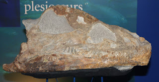
The skull of the "Shag Point Plesiosaur", now known as Kaiwhekea. That's pronounced "Ky-feh-key-uh"; one Maori pronunciation is "wh" as an 'f'.

The holotype skeleton of Kaiwhekea; yes folks, that's all one gigantic concretion that is ~20 feet long. It took a crew of 3-6 to collect those blocks over the course of a month (each day).

More photos will be coming soon!

Saturday, February 19, 2011

Trip to New York

New York City and Central Park from the 4th Floor of the
American Museum of Natural History.

A few weeks ago I took a trip to New York in order to be officially hired as a writer for a website on whale evolution that Jonathan Geisler (New York College of Osteopathic Medicine) is collaborating on with John Gatesy (UC Riverside) for an NSF grant. This trip was convenient because Jonathan and I are collaborating on a couple of research projects together, including the description of a fragmentary pilot-whale like skull from the Purisima Formation, as well as working on a large body of Herpetocetus material from Northern California. Once the website is up, I'll go into quite a bit more detail about it.

In 2008 I presented a talk (my first oral presentation) at SVP on a new skull of Herpetocetus bramblei I had found the previous year. The skull is pretty nice, and has about half the rostrum and a complete braincase, with earbones (tympanic, petrosal, stapes, incus, malleus). Because of its completeness and preservation, it will be a daunting task just to describe it. Fortunately (or unfortunately, depending upon how you look at it!) I have since collected another skull with a complete rostrum, and another (slightly less complete) skull from higher up in the Purisima Formation which may represent a younger species. So, including a fragmentary braincase at UCMP, there are four Herpetocetus crania, about a dozen petrosals, several tympanics, a half dozen dentaries, and some postcrania that we need to describe. Anyway, we didn't really work on this project at all.

One day during the trip we went in to the AMNH to look at some modern odontocete crania for comparison with the fossil pilot whale. A year and a half before, when I presented my poster on it at SVP (the 2009 Bristol meeting), I had identified it as Globicephala sp. at the time; fossil delphinid expert Giovanni Bianucci mentioned to me that he was not quite convinced that it belonged in that taxon. At the time I just identified it as best I could, without really having access to crania of other globicephalines like Pseudorca, Peponocephala, and Feresa. During our AMNH trip, we came to a similar conclusion as Giovanni - it's got too many differences to be in Globicephala, as it shares some other similarities with Pseudorca and Feresa.

Jonathan Geisler puzzles over fossil and modern globicephaline skulls.

However, it still definitely falls within the clade Globicephalinae, and that's what's important any way. I'll go into more detail about the ramifications of this fossil later on once we've at least submitted our paper - hopefully sometime early summer I'll have this one (manuscript #6) off.

Here I am (with appropriate shirt for the visit) with a beluga (Delphinapterus) skull.

Perhaps in a later post I'll post some of the pictures I took on a 15 minute dash through the fourth floor exhibits (which was all the time I had before we wanted to leave).


Wednesday, February 2, 2011

Mammal bite marks on fur seal bones, part 2

A few months after I collected the radius, I was invited to go examine Frank Perry's private collection. He's donated the majority of his material to UCMP, LACM, and the Santa Cruz Museum, but there was some remaining material. Several specimens he allowed me to borrow and prepare, including a partial juvenile Parapontoporia cranium, a walrus vertebra, and several fur seal bones. One of these was a very small humerus from a fur seal pup, roughly the same size individual as the radius I mentioned earlier. This specimen also happened to have a circular depression with a ring fracture, although it is much more shallow, and larger. This may be attributable to a larger, blunter tooth. Both of these specimens are probably attributable to the species Thalassoleon macnallyae, although in the article the bones are only identified to the family otariidae.
The right radius of a juvenile fur seal in anterior (left) and lateral (middle) views, and a closeup of the bite mark (right).

So, what caused these? Many, many, many studies have been published on shark tooth inflicted bite marks, which are typically linear gouges. These gouges are sometimes associated with removed 'chunks' of bone, but never have any fractures. These are obviously not linear gouges, and instead appear to be the result of the bone surface being pushed in. Circular holes can be caused by boring clams (pholad clams), but these are eroded, and do not result in fracturing. As it turns out, many similar tooth marks have been reported for conical mammalian teeth, of terrestrial mammalian predators and scavengers. One single similar tooth mark has been reported for a marine mammal: a skull of a juvenile sea lion (Eumetopias) from the Pleistocene of British Columbia (see the paper for more comments on this article). In fact, this is only the second reported occurrence of probable mammalian bite marks on fossil marine mammal bones.

Figure 2 from Boessenecker and Perry (2011) showing the bones and bone modifications.


The next question is, what type of mammal has the dental equipment capable of inflicting this sort of damage? Several pinnipeds, including the bizarre walrus Dusignathus santacruzensis, have teeth small enough to inflict these punctures. Most dolphins have teeth that are too small, and too closely spaced to make these punctures. Larger odontocetes, including the beluga relative Denebola, have larger teeth which are spaced far enough apart to form the punctures. Recently, Jonathan Geisler, Frank Perry, and I presented a poster on a pilot whale-like delphinid, and something the size of this cetacean could easily have produced the bite marks. The possibility remains that a terrestrial carnivore, like a canid, felid, or ursid; modern mammalian carnivores often prey upon or scavenge upon pinnipeds on shorelines. Lastly, the fur seal Thalassoleon has teeth that could produce the punctures. But Thalassoleon is the same species, you say! Well, oddly enough, extant fur seals and sea lions frequently commit infanticide - killing juveniles of their own species, sometimes in order to feed, other times as a part of aberrant sexual behavior where juveniles are mistaken for females.

Table of biogenic bone modifications from Boessenecker and Perry (2011) reported from marine vertebrate bones.

Unfortunately, it isn't possible to narrow the possibilities down any further. I'm getting tired, so stay tuned for part 3.

Tuesday, January 4, 2011

Odontocete skull excavation 2

Around 4 o'clock in the afternoon along the central California coast, the winter sun gets low on the horizon, giving a few minutes of lengthened shadows and rich, golden light, before it disappears behind a fog bank shortly afterward. The resulting decrease in light marks a premature sunset that can be barely workable sometimes. Winter field work is difficult even in California due to the shortened days, extreme tide fluctuations (even worse in Northern California), stormy weather, muddy trails, and low amount of sand on the beach (which results in a decrease in the number of access points and thus, fewer choices in access route). Oh, and not to mention poison oak, which during the winter, lacks its distinctive leaves). In this case, one of our access points was a small canyon which opens up towards the beach; during the winter, the stream fills the landward side of the canyon up with fetid, organic rich water while waves buildup a barrier bar, which dams the creek and forms a nice, disgusting little lagoon. When we arrived at this locality, the creek had pooled up, and was extremely cold as it had still been in the shade when we got there. The creek water hovered just over freezing, and was a good ten degrees colder than the ocean. It was still just as cold when we left; by the time we were done with the jacket, it was twilight. Oddly enough, on our way out, we came across a couple teenagers going down to the beach (the hard way, staying dry, but going up above the water-filled canyon on the side of a cliff) with only their cellphones for light. They must have made it out safely, although I'm not sure how (I never read in the paper about anyone dying out there).

Here's the excavated pit after the pedestal popped out.


Because the fossil-bearing pedestal had already popped off, we had to place the pedestal on a mound of sand to jacket it.

Chris takes burlap strips back to the excavation. I quite like this photo.

Just before we started the jacketing process. You can see in the background where the hole is, and that just a small sliver of the former 'fairweather' beach was left to stand on in order to reach the fossil. Within a few days, this was all gone. The sand surface you see there (where the red jacket is laying) is the higher surface of the beach from over the summer - just a small remnant of it remains. After this sliver is gone, the fossil would have been many feet out of reach above the beach.

A beautiful sunset (and advancing rainclouds) heralded our completion of the excavation.

The odontocete skull is halfway jacketed, and as the temperature begins to drop, our hands were beginning to go numb.

Chris cuts more burlap strips for the second half of the jacket.

Finally! Just before dark, the plaster jacket is completed, and we're ready to lug all the gear and fossils back up the trail.

Monday, January 3, 2011

Odontocete skull excavation 1

Over Thanksgiving break in 2009, while scouting out a locality for a future permit application, I spotted a nice braincase of a small odontocete skull exposed in a cliff face. The following november, I received a permit, and returned to the locality one year later with my friend Chris Pirrone to excavate the fossil. I knew this excavation would be slightly challenging, because we would be pedestaling the fossil sideways instead of from the top (I have done worse before - I have successfully trenched and pedestaled fossils in overhangs, including an odontocete skull).


Chris excavating the cranium.


Chris excavating later in the morning.


The skull by midafternoon.

By the middle of the afternoon, we had excavated a ring shaped hole around a cylinder of rock which contains the skull. A few pieces broken off (and glued back on) indicated that the skull was very well preserved. However, given the remaining amount of rock, I was concerned that we would not be able to finish before 5pm. Additionally, while excavating on the left side of the hole, I split off a piece of rock which contained a fragment of the rostrum. This indicated the rostrum was relatively long - a scary prospect, perhaps meaning I would have to return and finish the excavation the following day.


Here's the posterior braincase exposed: the right side of the skull is exposed, and the skull is upside down. The right squamosal, and occipital condyle are clearly visible in this photo.

After continued trenching around the cylinder of rock, at about 4pm, the base of the pedestal snapped unexpectedly, and the whole thing came out in one fifty pound piece. The anterior end of the pedestal terminated against an oblique fracture surface; I was nervous that the rostrum may have continued through this fracture. I carved off a half-inch from this surface, and found no bone; additionally, I found no bone in the end of the pedestal. Fortunately, this means the entire skull is preserved within the block.

Saturday, January 1, 2011

Associated dolphin vertebrae

Sorry for the delay in posting; I've been enjoying a lazy winter break so far. Over thanksgiving break, I spent some time at a Purisima Formation locality which I had just received a new collections permit for. I went out with my friend Chris Pirrone to this new locality. Unfortunately, there hadn't been much storm activity since the summer. Storm activity achieves two things: it washes off a ridnd of weathered clay minerals off (in addition to algae), and it transports sand from the beach to offshore bars, which during fairweather, are slowly eroded and the sand is transported back onshore.
After a while of not finding anything, I spotted these three associated vertebrae - from some kind of a small odontocete. Two of the vertebrae lie in near articulation, and the other is slightly displaced.


Here's a closeup view of the specimens.


Chris Pirrone applies vinac to the fossil vertebrae.

We collected these three vertebrae, but it was too cold to stay and dig through the sediment for more bones. I'm sure there are more; some forelimb or cranial elements would be great. I'll return to the locality once some more storm activity cleans off the cliff exposures.

During the course of my taphonomic research of fossil vertebrates in the Purisima Formation, I've found that associated vertebrate remains (two or more elements which in life are only joined by soft tissue) are extremely rare in the shallow marine fossil record (if the Purisima Formation is taken to be a representative shallow marine deposit). Indeed, this is one of a couple dozen specimens showing any degree of association or articulation.

Friday, November 12, 2010

Shark-bitten dolphin skull

In 2008 I spent the day before Christmas Eve shivered on a cold, wind-blasted California beach prospecting for vertebrate fossils in the Purisima Formation. I was home on winter break, and although it is far more cold where I go to graduate school in Montana (as I write this I'm looking out at the results of our first winter snow), nothing is worse than being wet and miserably cold out on the foggy, windy coast of the golden state (except perhaps being wet and miserable on the Oregon coast, which I've done).

The thrill (or promise) of discovery is more than enough to keep me fueled in the field during the winter. Indeed, when the birds start singing and the snow melts in the spring, most paleontologists start to get field fever - the field season for most vertebrate paleontologists is during the summer months. Anyone who's ever tried to do coastal fieldwork during the summer, on the other hand, is in for a rude awakening. No erosion takes place during the summer, and many of the outcrops are totally buried. The exposures that are above the beach sand level (which is higher during the summer) are typically covered with dust, sand, and grime, which obscures fossils. The storms in the winter months clean this nasty coating off, and transport beach sand into offshore bars, often exposing strata below the beach (I see new fossil localities every winter this way). Winter is my field season.

Historically, I've had really good luck the day before Christmas Eve. It's my last day before Christmas to make it out in the field. The prior year, I found a humongous Carcharocles megalodon tooth (the only specimen known from the Purisima Formation), and discovered a partially articulated fur seal skeleton.

The Christmas Eve dolphin.

At 4pm, the tide was beginning to come back in, and with little over an hour of daylight, it was looking like I was going to come home empty-handed. I went to one last cove before I turned around to head back to the beach. I walked for a few minutes and spotted something in a boulder I had not seen on my way out: a pair of flat bones joined along an articulation that looked suspiciously (even from 20 feet away) like the palate of a dolphin skull. Upon closer examination, yes indeed! It was a dolphin skull in a mollusk shell bed; the width and flatness of the palate suggested it was not Parapontoporia, the most common odontocete in the Purisima Formation. I set about chopping into the boulder; fortunately, most of it was relatively soft. However, an extremely hard calcium-carbonate cemented concretion the size of a basketball had formed over the dorsal surface of the braincase and rostrum, and this slowed digging down. By dusk, the concretion didn't budge. After another half hour, it finally popped out of the boulder, and I lugged the 45 pound block back to my car. Exhausted, I drove home, drank a couple of hard-earned beers with dinner, and passed out.

View of the facial region of the skull.

When it came time to go back to Montana, I decided I would rather take the fossil as a carry-on than risk checking it and picking up a broken fossil that I had paid 25 bucks for thanks to baggage fees. After arriving in Bozeman (with a very sore back and neck from lugging 65 pounds
of luggage through the Denver airport), I almost immediately began preparation (starting, of course, with acetic acid baths for several weeks to soften the concretionary matrix). It took about two months to prepare, and as you can see from the above photos, it is damn beautiful. I initally identified it as something like Haborophocoena - it bears numerous similarities. However, after showing them photos of the specimen at SVP 2009 in Bristol, UK, Olivier Lambert and Giovanni Bianucci both think this represents a basal delphinid rather than a basal phocoenid. I'm inclined to agree with them, although part of my original ID was based on the presence of premaxillary eminences, which this specimen has (a phocoenid character). However, the ascending process of the right premaxilla is in contact with the nasals while the left is not (a delphinid character). Whatever it is, it will require preparation of the ventral aspect, and more careful analysis of the morphology than what I've been able to do thus far. Whatever it is, it appears to represent a new genus and species, and will make a beautiful holotype specimen in the future. During preparation, one curious thing I noticed was a notch in one of the premaxillary eminences (the large pads/bumps in front of the bony nares). I initially dismissed it as a pathology.

The left premaxillary eminence showing linear gouges (red lines) and missing bone.

Upon closer examination (which admittedly did not occur until yesterday, almost two years after collection) it became apparent that the abnormal area had two distinct, paralell linear gouges, and a short, less distinct third one in the middle (this one is still partly filled with matrix). Around these gouges is an area of exposed cancellous bone, where the bone has been removed.

Additional gouges present near the base of the rostrum.

I also found four more gouges present: two long ones, and two short ones; all but one are parallel. In fact, aside from the one gouge seen above trending towards the upper left corner of the photo, all the gouges are parallel. This is a textbook set of shark-inflicted bite marks. There are a lot of papers on this in the literature, documenting shark bites on dolphins, baleen whales, pinnipeds, sea turtles, other shark teeth, mosasaurs, plesiosaurs, dinosaur bones, sea stars, and probably other marine critters as well.

In fact, the first record of these types of trace fossils were actually first documented in the modern environment: on predated and scavenged sea-otter carcasses from Monterey Bay, and reported by Ames and Morejohn (1980). The reported linear gouges, subparallel wavy small gouges, and a specimen including a shark tooth embedded in a sea otter skull. The morphology of the traces along with the tooth identified the culprit as the Great White Shark, Carcharodon carcharias. Two years later, these exact types of traces were identified by Tom Demere and Richard Cerutti (1982) on a baleen whale dentary (of my favorite whale, Herpetocetus!), and identified as "Carcharodon sulcidens" (a taxon now just considered to be fossil Carcharodon carcharias).

It's not clear what type of shark fed on my poor little dolphin, or if it was a case of predation or scavening; from what I've read, the majority of carcasses that exhibit bites have bite marks on the posterior portion of the body, which is just about as far as you can get from the face. This makes total sense, given how a shark would have to bite into a fleeing dolphin during pursuit. Furthermore, it's interesting to note that this bite would have had to go clean through the dolphin's melon (if it had not already decomposed). Anyway, I interpret these traces as drag marks from the apices of the shark's teeth; I suppose later on I can figure out the relative motion of the shark's mouth during the bite (most likely lateral shake feeding). It'll make for a nice short paper some day...

Ames, J. A., and Morejohn, G.V., 1980, Evidence of white shark, Carcharodon carcharius, attacks on sea otters, Enhydra lutris: California Fish and Game, v. 66, p. 196-209.

Deméré, T.A., and Cerutti, R.A., 1982, A Pliocene shark attack on a cetotheriid whale: Journal of Paleontology, v. 56, p. 1480-1482

Tuesday, November 2, 2010

A bizarre new pilot-whale relative from the Pliocene of the North Sea

This morning when I woke up I had a nice message on facebook from a friend of mine - a link to a press release on a new fossil dolphin from the North Sea. To be honest (I had just woken up and was still fairly groggy when I first read it) I first saw this beautiful painting below, and not quite realizing it was a painting, the first thing I thought was "whoa, another extant cetacean???"

I then thought, "whoa, that reconstruction looks really weird."

Reconstruction of Platalearostrum hoekmani from Post and Kompanje (2010) by Remie Bakker.

Fortunately, I was easily able to find a link to the pdf for the article (which came out today in the European journal Deinsea) and see why the beast in the painting looks so strange. Post and Kompanje (2010) report on a fragmentary cranium of a bizarre new odontocete. While very incomplete, the preserved portion of the rostrum (="snout") does exhibit some strange features - a very low tooth count (6 sockets/alveoli), a laterally convex toothrow, and an insanely wide lateral 'wing' of the premaxilla, which makes the rostrum wider towards the anterior tip. The maxilla is also not the lateral most portion of the rostrum towards the front - another weird feature (which is shared in the pilot whales, Globicephala, and the extinct Protoglobicephala). The lateral 'wing' of the premaxilla is also pointed 'upwards' (dorsally) a bit, to make the dorsal rostrum surface concave, like a giant spoon. Although totally weird, the rostrum shares a number of features with the pilot whale Globicephala - anteriorly widening premaxillae, rugose bone surface on the premax, a short laterally convex toothrow, and a rostrum that is pretty short and blunt in general. Globicephala in general is already a very strange critter, and Platalearostrum makes it look sober in comparison.


The holotype fossil of Platalearostrum hoekmani (modified from Post and Kompanje, 2010)

Other features (aside from its close affinity with Globicephala) indicate its inclusion within the clade Globicephalinae. The authors curiously chose to use the clade Orcininae - in the usage of Bianucci (2005). Recent molecular analyses have shown the Globicephalinae paraphyletic in the sense that Orcinus is usually not included - and everything else (Globicephala, Feresa, Grampus, Pseudorca, Orcaella) make a monophyletic group. Without Orcinus, it just isn't the Orcininae anymore. Orcininae may be a valid term if fossil taxa like Hemisyntrachelus and Arimidelphis are shown to be sister taxa of Orcinus orca and the extinct Orcinus citoniensis. That being said, Globicephala is not in that group. Which is all the more interesting, suggesting two different clades of delphinids trending toward (relative) gigantism during the Pliocene.


Comparison of Globicephala macrorhynchus and Platalearostrum hoekmani (from Post and Kompanje, 2010)

This article made me pretty happy because I've worked a little on fossil globicephalines from the Pliocene of California. There aren't that many - bona fide delphinid fossils are generally quite rare in the Mio-Pliocene record in California, as opposed to the obscenely delphinid rich (and diverse!) Pliocene fossil record of Italy. Things like Orcinus, Globicephala, Protoglobicephala, Arimidelphis, and Hemisyntrachelus are all already living (not necessarily coexisting) in the world's oceans in the Pliocene, and now we have another weird one on top of this. Certainly it can be said that during the Pliocene, delphinids were experimenting with new "body plans" (loosely using the term) and rapidly diversifying. The Pliocene was a weird time, and boasted a combination of many strange marine mammals which were more derived than extant relatives often with novel anatomical features and adaptations, relatives of modern taxa with wider geographic distributions, and holdovers of archaic taxa which had not yet kicked the proverbial bucket.

Klaas Post, Erwin J.O. Kompanje, 2010. A new dolphin (Cetacea, Delphinidae) from the Plio-Pleistocene of the North Sea. Deinsea 14:1-14

Bianucci, G., 2005. Armidelphis sorbinii a new small killer whale-like dolphin from the Pliocene of the Marecchia river (central eastern Italy) and a phylogenetic analysis of the Orcininae (Cetacea: Odontoceti) - Rivista Italiana di Paleontologia et Stratigrafia 111: 329-344

Wednesday, January 20, 2010

Uranocetus and hearing in mysticetes

Hey Folks, Sorry about the delay; I realize its been over a month since I last posted anything. Winter break was not exactly relaxing, and the parts that neared relaxation were spent doing fieldwork (which definitely yielded some interesting material). In other news, my first technical paper has been tentatively accepted for publication by the UCMP-published journal PaleoBios; I'm approximately 99% done with revisions at this point, so you'll hear more about it after it's in press.

Recently two mysticete related papers have been published - Erich Fitzgerald's monograph on the truly bizarre Mammalodon colliveri, which I'll cover later, and M.E. Steeman's (2009) thought provoking paper naming the new "cetothere" Uranocetus from the Miocene of Denmark and its implications for mysticete hearing.

The cranium of Uranocetus, from Steeman (2009).


First off, "cetotheres" are a wastebasket group of generalized archaic baleen whales that don't fit nicely in modern families, although Bouetel and Muizon (2006) have redefined the Cetotheriidae sensu stricto as a small group with some very strange cranial features, including my personal favorite, Herpetocetus. Most other cetotheres (Cetotheriidae sensu lato) were placed into newly named families (Pelocetidae, Aglaocetidae, and Diorocetidae) which were sister taxa to the Balaenopteridae all included in her concept of the Balaenopteroidea (but not in the concept of the Balaenopteroidea advocated by Demere et al. 2005, which is Eschrictiidae + Balaenopteridae). Bottom line is Uranocetus is some kind of stem baleen-bearing mysticete, no matter whose phylogeny you use. The dentary of Uranocetus (from Steeman, 2009).
Interestingly, while it is placed rather close to Balaenopteridae, it still retains a large mandibular foramen, a plesiomorphic feature for mysticetes. The mandibular foramen is very small in extant mysticetes, but extremely large in odontocetes, so much that the posterior portion of the dentary is a thin bony shell (the "pan bone") that houses the mandibular fat pad. The lateral margin of the dentary is extremely thin, so that high frequency sounds can pass through without significant volume loss (Nummela et al. 2007, Steeman 2009). High and mid frequency sounds pass through this, and are then channeled up through the mandibular fat pad and up to the tympanic plate; in odontocetes, this is more or less a functional analog of the external ear pinna. And, by the way, all these strange auditory features are adaptations for allowing directional hearing underwater; otherwise terrestrial mammals hear via bone conduction hearing (sound travels faster in water, and the mammalian body is roughly as dense as the surrounding aqueous medium), and sounds more or less arrive at each ear too quickly to discern the direction. Cetaceans have separated their ear bones (petrosal, tympanic, and ossicles) from the temporal bone and surrounded them by sinuses to isolate these complexes from the skull to hear directionally. While this was initially thought to be an adaptation for hearing high frequency sounds and thus an adaptation for echolocation (a capability restricted to the odontoceti, and associated with high frequency sounds), recent research has identified the pan bone/enlarged mandibular foramen (i.e. bony correlates of the mandibular fat pad) in many archaeocetes, including Ambulocetus, remingtonocetids, protocetids, and basilosaurids (Nummela et al. 2007) as well as many archaic toothed- and toothless mysticetes, such as Aetiocetus weltoni, Mammalodon, Eomysticetus, and even Herpetocetus. This led Nummela et al. (2007) to reason that, since neither archaeocetes or mysticetes have any anatomical features associated with echolocation, that this feature is probably instead related to underwater hearing in general, and not just echolocation.
Dentaries of various archaic mysticetes and an archaeocete, from Fitzgerald (2009).

The fact that most basal mysticetes have an enlarged mandibular foramen suggests that this is a feature inherited from basilosaurid ancestors. Interestingly, modern mysticetes are adapted for hearing low frequency sounds, which pass through dense bone without significant volume loss. While Uranocetus has a large mandibular foramen, the lateral wall is too thick to be useful for hearing anything aside from low frequency sounds (which Uranocetus is adapted to hear based on its cochlear structure; Steeman 2009). The exact same thing is seen in Herpetocetus, which is also adapted for low frequency hearing, but has a large foramen with a thick lateral wall. This suggests that at least in these later diverging taxa, that the large mandibular foramen was a vestigial feature perpetuated by phylogenetic inertia.
Lateral aspect of a (not so typical) mysticete (Eshrichtius robustus, the Gray Whale) skull and dentary in articulation, from Johnston et al. (2009).


Steeman (2009) reasoned that the mandibular foramen decreased in size to strengthen the dentary due to the intense forces involved during feeding. Above shows a gray whale skull and mandible in articulation, just to give you an idea of how strange the mysticete feeding apparatus is (exclusive of baleen). In any event, I've been thinking about this quite a bit recently, and got to add a (very short) synopsis of this in my manuscript revisions, but you'll hear about that soon enough.

References-
Deméré, T.A. and A. Berta (2008). Cranial anatomy of the toothed mysticete Aetiocetus weltoni and its implications for aetiocetid phylogeny. Zoological Journal of Linnean Society, 154(2): 308-352. PDF

Deméré, T.A., A. Berta, and M.R. McGowen. 2005. The taxonomic and evolutionary history of fossil and modern balaenopteroid mysticetes. Journal of Mammalian Evolution 12:99-143.

Fitzgerald, E.M.G. 2009. The morphology and systematics of Mammalodon colliveri (Cetacea:Mysticeti), a toothed mysticete from the Oligocene of Australia. Zoological Journal of the Linnean Society 110p.

Johnston, C., T. Deméré, A. Berta, J. St. Leger and J. Yonas. 2009. Observations on the musculoskeletal anatomy of the head of a neonate gray whale (Eschrichtius robustus). Marine Mammal Science PDF

Nummela, S., J.G.M. Thewissen, S. Bajpai, T. Hussain, and K. Kumar. 2007. Sound transmision in archaic and modern whales: anatomical adaptations for underwater hearing. Anatomical Record 290:716-733.

Steeman, M.E. 2009. A new baleen whale from the late Miocene of Denmark and early mysticete hearing. Palaeontology 52 :1169-1190.

Sunday, December 6, 2009

Fossil preparation - odontocete tympanic

About two or three summers ago I collected a beautiful little odontocete tympanic from the Purisima. Problem was, I only found out it was beautiful (past tense) after it sat in about twenty or thirty pieces. Because the part that was exposed looked like some other type of bone (and not an odontocete tympanic) I mis-estimated how sturdy the fossil was, and it exploded as I carved matrix away from it. I have since not repeated the mistake. Anyway, the fossil has sat in pieces in a plastic bag for two years, and I finally got the courage to glue it back together. I say courage because 1) I was somewhat embarassed by this damage, and 2) I was nervous to piece together all these tiny fragments. Below is a photo of what I had to work with.
Tympanic fragments prior to preparation.

I began by finding the pieces of the robust involucrum, which is the thick portion of the cetacean tympanic. There are more or less three major portions of the tympanic: the involucrum (frequently the only preserved part), the posterior process, which attaches to the posterior involucrum, and the paper-thin involucrum (which in odontocetes is usually under 1-1.5mm thick, hence the overall fragility of these elements). Then, I started finding matching pieces, and gluing these to the involucrum.

The tympanic after 30 minutes of preparation; some of the outer lip fragments have been glued in place. Lateral view (top photo) and anterior view (bottom photo) - note the matrix-free tympanic cavity.

After a couple more hours, I was able to finish gluing back most of the outer lip of the tympanic, as well as the posterior process.

Tympanic before (left) and after (right) addition of the sigmoid and posterior process.

Tympanic in dorsal (left) and lateral (right) aspects.

After the fossil was glued together, it became very obvious that this was a tympanic from the "river dolphin" Parapontoporia wilsoni, which has a small posterior process, a sharp anterior apex of the bulla, and most characteristically a laterally inflated outer lip, not seen in any other Purisima odontocete (for which tympanics are known, and out of the given tympanic sample from the Purisima Fm.). As far as crania, jaws, periotics, tympanics, and parts thereof go, Parapontoporia is by far the most common Purisima odontocete (i.e. between collections at UCMP, SCMNH, and LACM go, there are roughly a dozen nearly complete crania known, rostrum not included).

Newly prepared tympanic (right) side by side with another very well preserved Parapontoporia wilsoni tympanic.

All in all, I was extremely pleased; in one afternoon I had turned a pile of fragments (which I had virtually no hope for) into a beautiful little specimen. All but three tiny fragments under 5mm in size were glued on; the other ones probably attached to the margin of the outer lip, which may require fragments lost during collection (or, conversely, pieces pulverized). But let's not split hairs here - this by far was the most damage I've ever done to an odontocete tympanic, to the point where I was embarassed to even think about it; and now, it's one of the nicest I have. The positive side to this inadvertently destructive mode of
collection was that all the matrix was absent from the tympanic cavity, unlike the specimen on the left in the above photo (where the matrix inside was actually phosphatized, but phosphatic 'cementation' had fortuitously not formed an overgrowth around the rest of the bulla - best case scenario!). This is one of about a dozen and a half or so odontocete bullae I've recovered from the Purisima.

Monday, November 2, 2009

New lower jaw of the extinct lipotid Parapontoporia

Before I dive back into fossil odobenids, I'd like to show off something I just finished preparing. I collected this specimen from underwater in July in Santa Cruz County; I arrived at the exposure several days after I initially discovered it and coated it with vinac. Unfortunately, upon my return when I intended to collect it, the tide wasn't low enough; the specimen was exposed on the apron of a cliff, and was about 6" underwater. Unfortunately, I was there at low tide, and in the intervening days, the sand on the beach had locally eroded, allowing waves to go *just* a bit higher on this 5m stretch of beach.

Oblique view of the lower jaw of Parapontoporia wilsoni.

Anyway, after forty minutes of carving out a pedestal, cursing like mad because I thought I was going to destroy the fossil, and being investigated (and probably secretly laughed at) by a sea otter and a sea lion, I decided to undercut the pedestal. The pedestal was about 14" long and 5" wide, and I was worried that it might crack in half during undercutting - any bone exposed in that crack might fall out (and be swept away by the surf), and then I wouldn't be able to connect the bone from the two pieces of the pedestal back together. Needless to say I was shocked (and endlessly pleased) when the pedestal popped off perfectly.

Dorsal aspect of the fused dentaries of Parapontoporia wilsoni.

Lateral aspect of the fused dentaries of Parapontoporia wilsoni.

Parapontoporia is a very conspicuous member of late Neogene marine vertebrate assemblages in California and Baja California, and has also been reported from Japan. In California, it is known from late Miocene (Tortonian - 9 Ma) through late Pliocene (Piacenzian - 2 Ma) strata, including the San Mateo, San Diego, Capistrano, Pismo , Purisima , and Wilson Grove Formations. Three species are known - Parapontoporia pacifica from the late Miocene Almejas Formation of Baja (Barnes, 1984), Parapontoporia wilsoni from the Mio-Pliocene Purisima Formation (Barnes, 1985), and Parapontoporia sternbergi from the San Diego Formation (Barnes, 1985).

The cranium and jaws of Parapontoporia sternbergi, on display at the San Diego Natural History Museum.
Since description, P. pacifica is still only known from one partial cranium, while P. sternbergi is now represented by about a dozen well preserved crania, and several nearly complete lower jaws. While only the partial holotypic cranium of Parapontoporia wilsoni has been described, however, there are now probably around two dozen crania known, in addition to around 50-75 periotics. Only two lower jaws are known, though - one crappy fragment at UCMP, and a neat (but highly abraded) fragment of an articulated rostrum with teeth at CAS. Two more well preserved jaws are known, both from the early Pliocene of the Purisima - one I collected with my girlfriend in 2006, and the specimen I collected this summer. The 2006 specimen has one tooth, but is better preserved than this specimen.
This specimen may not represent P. wilsoni; the P. wilsoni holotype is about a million years older, and it is certainly possible that crania from this stratum represent P. sternbergi due to their younger age; description of material from the San Mateo Formation is needed to investigate this further. In fact, a huge body of fossils of Parapontoporia need to be described.
Closeup of the teeth of the new jaw of Parapontoporia wilsoni.

Parapontoporia was originally named for its similarity to the extant La Plata River dolphin, or Franciscana (Pontoporia blainvillei; Barnes, 1984, 1985). However, subsequent studies have placed it within the Lipotidae, as the sister taxon of the now extinct Yangtze River Dolphin (Lipotes vexillifer; Geisler and Sanders, 2003; Muizon, 1988), which was only described in 1918. Parapontoporia has an extremely long rostrum and mandibular symphysis, and *may* have the most teeth of any mammal (which, if it isn't Parapontoporia, I'm sure it's some kind of eurhinodelphid or other longirostrine odontocete from the Chesapeake Group of the east coast).

Wherever Parapontoporia occurs, it dominates the odontocete assemblage - in the Purisima, up to 38% of isolated periotics are referable to Parapontoporia. The most abundantly known odontocete crania from the Purisima belong to this taxon. Interestingly, despite decades of construction in San Diego, there are now more crania of this taxon known from the Purisima than from the San Diego Formation. Many of these Purisima crania are still in concretions, but nonetheless, they exist, and an excellent opportunity for a study of ontogenetic and stratigraphic variation is possible given this sample (a project Nick Pyenson was bugging me to do, but I simply didn't have the time as an undergrad). In fact, I picked up two partial crania this summer (both in nodules, though; one weighed about 55 pounds).

Nick Pyenson (2009) recently published a pretty neat (albeit depressing) paper in marine mammal science about the consequences of the extinction of Lipotes, given its 'colorful' evolutionary history. But this post is long enough as is, and I could do several more posts just on Parapontoporia; I'll save discussion of that paper for later.

BARNES, L. G. 1984. Fossil odontocetes (Mammalia: Cetacea) from the Almejas Formation, Isla Cedros, Mexico. Paleobios 42:1–46.
BARNES, L. G. 1985. Fossil pontoporiid dolphins (Mammalia: Cetacea) from the Pacific coast of North America. Contributions in Science, Natural History Museum of Los Angeles County 363:1–34.
GEISLER, J. H., AND A. E. SANDERS. 2003. Morphological evidence for the phylogeny of Cetacea. Journal of Mammalian Evolution 10:23–129.
MUIZON, C. de. 1988. Les relations phylog`en´etiques des Delphinida (Cetacea, Mammalia). Annales de Paleontologie 74:159–227.
PYENSON, N.D. 2009. Requiem for Lipotes: an evolutionary perspective on marine mammal extinction. Marine Mammal Science 25:714-724.