Wednesday, April 19, 2023

New fossils of Coronodon 2: two new species of toothed mysticetes from South Carolina

 This is a continuation of my blog 'coverage' of our new monograph - for an introduction to the big new paper, see the first post in this series here. For the third post in this series, click here.

But first - it's been a pretty dark day for me and Sarah, so I need to share some memes that were made out of/inspired by my artwork:


Family portrait of Coronodon that I illustrated last year and finished in February: upper left, juvenile Coronodon havensteini; lower left, adult Coronodon havensteini; upper right, Coronodon planifrons; lower right, Coronodon newtonorum. Graphite and digital, 2023. Original about 10x14".

My illustration has inspired some pretty fantastic memes - my artwork does look like the other Coronodon whales are laughing at the one. Cartoon by M. Nur Yahya on twitter.


A variation of an older meme - by @natespithe on Twitter.

One of my favorites - by Henry Tsai on Facebook.

And lastly, the Coronodon whales cartoonized by @Yoofilos on Twitter into the "Me and the Boys" meme from above.

Stratigraphic Distribution of Coronodon

I'll dive into the rationale for naming new species shortly, but first I'd like to discuss the stratigraphic distribution of specimens. The holotype skull of Coronodon havensteini was collected from a submerged exposure of the Ashley Formation - a late early Oligocene sandy limestone. It's firm, with some concretions here and there, generally kind of a brownish tan to pale yellow color and occasionally can be olive green or khaki - frequently fossiliferous, with sparse mollusks, barnacles, and bryozoans. The Ashley Formation has been dated with Strontium isotopes to about 28-30 Ma - uppermost Rupelian, which is the stage occupying the first half of the Oligocene epoch. All new specimens we referred to Coronodon havensteini are also from the Ashley Formation. The two specimens that ended up being holotypes of new species - CCNHM 166 and ChM PV 2778 - were both collected from the Chandler Bridge Formation. When Al Sanders and others named the Chandler Bridge Formation in 1986, they remarked that the whale faunas were so similar that they were likely only separated in time by about a million years or so. Somewhat surprisingly, strontium isotope dates indicate that there's about 4-5 million years - and that the Chandler Bridge Formation is 24.7-23.5 Ma or so (Weems et al., 2016). Point is - these Coronodon specimens are separated by a bit of time. There are some other subtle differences: eomysticetids seem to be stratigraphically separated, with all specimens of Micromysticetus being derived from the Ashley and all specimens of Eomysticetus originating from the Chandler Bridge. Xenorophid odontocetes appear somewhat stratigraphically separated: more plesiomorphic taxa, like Xenorophus and Albertocetus, make up most of the xenorophid assemblage from the Ashley whereas there are additional, more derived taxa in the Chandler Bridge including Cotylocara and Echovenator, as well as at least one additional Cotylocara-like genus that has not yet been named. There are perhaps a half dozen waipatiids in the Chandler Bridge, but only two or three forms from the Ashley, and they seem to be restricted: a dwarf form, and Ediscetus osbornei named a few years ago. Another species of Ediscetus may be present in the Ashley - and that glut of waipatiid specimens will be my major project this summer. 

Stratigraphic distribution of Coronodonidae in the Oligocene rocks of Charleston, South Carolina.


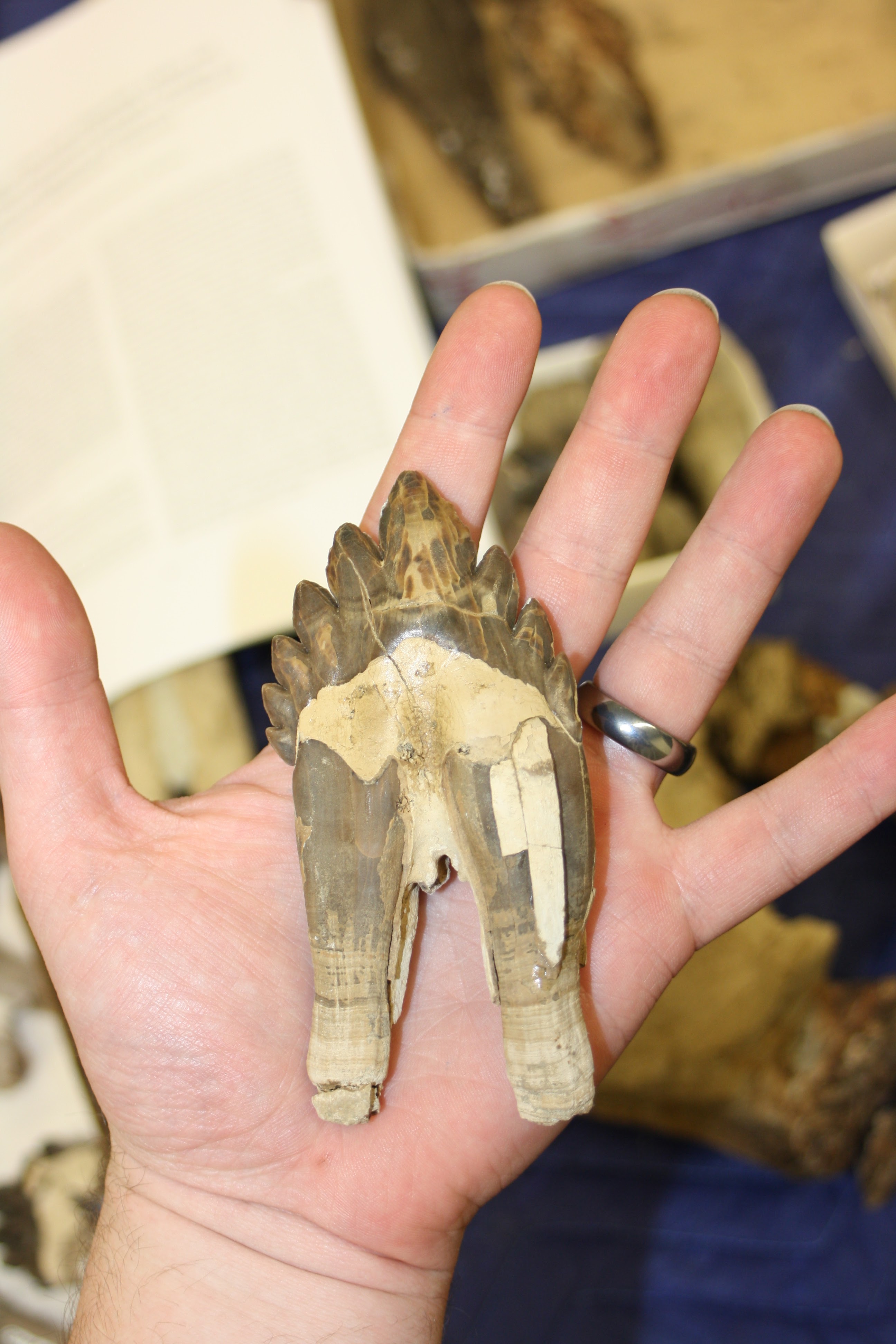
 The holotype skull of
Coronodon planifrons, CCNHM 166, the Volcko-Chapman whale.

Recognizing the Existence of New Species of Coronodon

Discovering a new species - whether you're digging it up and doing all the sweaty work, or the actual anatomist doing the descriptive work - is not always obvious. For a long time I thought there was a single species - after all, I've already reported (or interpreted) both of the odontocetes Albertocetus meffordorum and Ankylorhiza tiedemani as being present in the Ashley and Chandler Bridge formations. My working hypothesis was "one species in both units" - which I feel, from the perspective of a lumper, the most conservative approach. When I arrived at CCNHM in 2015, the specimen that is now the holotype of Coronodon planifrons, the "Volcko whale" CCNHM 166, and the "Coosaw whale" CCNHM 164, were both in our collections but not included in the original study. The "Volcko whale" was found in 2010 and the Coosaw whale a few years earlier (each within a couple miles of our house!). I sat down and looked for differences earlier on back in 2018 and actually failed to make a number of observations I made later - but I did make one important observation back then: the holotype specimen of Coronodon havensteini, and what is now the holotype of Coronodon planifrons (CCNHM 166), had differently shaped supraorbital processes of the frontal. There are these little processes on either side of the eye socket, helpfully named the preorbital and postorbital process (measuring between them, by the way, gives you eye socket size). In Coronodon havensteini these processes are the same thickness if you measure from the inside out. However, in CCNHM 166, the postorbital process is quite a bit thicker than the preorbital process (and vice versa). Now, these bones are also very porous so I thought, maybe this is just caused by wear to the skull, and discounted species-level differences.

A comparison of the preorbital and postorbital processes of the three species of Coronodon - some of the earliest observations I made. Coronodon havensteini has pre- and postorbital processes of equal depth, Coronodon planifrons has a deeper postorbital process, and Coronodon newtonorum has a deeper preorbital process.

About a year ago I noticed that the supraorbital process of the frontal, in anterior view, was nearly horizontal - and I only noticed this because I but the CCNHM 166 skull, along with referred C. havensteini specimen CCNHM 164, backwards, with the posterior end of the skull facing the wall. In both CCNHM 164 and the holotype CCNHM 108, the supraorbital processes descend at an angle and are decidedly not horizontal. I think I made that observation during a meeting. So, I pulled the specimens out and started more detailed comparisons. I noticed that the sagittal crest was much longer in CCNHM 166, and that the frontal-parietal suture is much more V-shaped. I also noticed that the sternomastoid fossa - a D-shaped rugose pit for the sternomastoid muscle, which is positioned on the squamosal bone and just above the jaw joint - has this deep trough that rises up along the side of the nuchal crest; this is missing in Coronodon havensteini. At this point I was more or less convinced that the number of anatomical differences had exceeded individual variation for Coronodon havensteini and instead suggested to me that CCNHM 166 represented a different species.

 
The holotype skull and mandible of Coronodon planifrons, CCNHM 166 - the Volcko-Chapman whale.

A parallel development happened in Fall 2017 after the first paper was published - collector Taffie Chapman, who found parts of CCNHM 166 in the field, donated specimens she had first found and kept. A bunch of missing parts clicked back together - for example, the condyle of the mandible was in her material! Taffie kept one tooth for herself, a nice premolar, and I did ask for it - but she politely insisted, and I knew better than to push it. Included amongst her material were several more vertebrae and a bunch of partial teeth. As a result, we now call it the "Volcko-Chapman whale". Years later, last summer actually, Jonathan Geisler and I finally got a chance to compare these side by side and we realized several teeth from the original sample were improperly glued together - and, within 20 minutes of when Geisler was supposed to drive back to New York after a vacation here in Charleston. I airscribed away some putty in the prep lab and texted him photos of the preliminarily reassembled teeth - he texted back something along the lines of "this changes quite a bit."

 The periotics (ear bones) and dentition of Coronodon planifrons, the Volcko-Chapman whale.

One of the first changes were the smallest (and furthest posterior) molars - the smallest one had broken in half, one half found and kept by each collector. I glued them back together, revealing a tiny upper third molar (M3) that was approximately 2/3 the diameter of the second to last tooth (upper second molar, M2). The M3 is unknown in the holotype, but the upper M2 is preserved, and the "Coosaw whale" specimen of Coronodon havensteini (CCNHM 164) has both the M2 and M3. These teeth are of similar size in Coronodon havensteini, and also larger than both teeth in CCNHM 166 (Coronodon planifrons). In other words, CCNHM 166 "Volcko-Chapman whale" (Coronodon planifrons) has 1) smaller molars and 2) molars that decrease in size posteriorly. We initially thought there were some differences in cusp counts, but that changed when we settled upon the final identification of teeth to position after discovering the tiny size of the M3 - after this, the cusp counts were quite similar. I took another look at the earbones, and found that there were also a few features of the periotic bone that were unique - for example, the lateral tuberosity extends far laterally, whereas it is a small bump in the holotype of Coronodon havensteini. All of these features ended up in our diagnosis* of Coronodon planifrons. The tiny size of the upper M3 also permitted us to refer an isolated molar to Coronodon planifrons, also showcasing what the tooth shape would be without any tooth wear. I suggested naming the species after its most distinctive feature: horizontal frontal bones over the orbit, hence Coronodon planifrons.

*The first time I read a scientific paper and saw diagnosis for a species I thought "what the hell? A diagnosis is for a disease" but as it turns out, a diagnosis in zoology and botany (or taxonomy in general) is a list of the different anatomical/morphological features that are used to identify specimens of that species. In a sense, it is one of the most important parts of the reporting of a new species, genus, or family. Ewan Fordyce impressed upon me how important it is to craft a useful, lasting diagnosis that future researchers can use. One pet peeve of mine is the lazy use of synapomorphies from a cladistic analysis and restricting your diagnosis to just features from the cladistic character list: these diagnoses are usually not very useful. Medical diagnoses are constructed similarly: a suite of diagnostic features used to identify a medical condition.


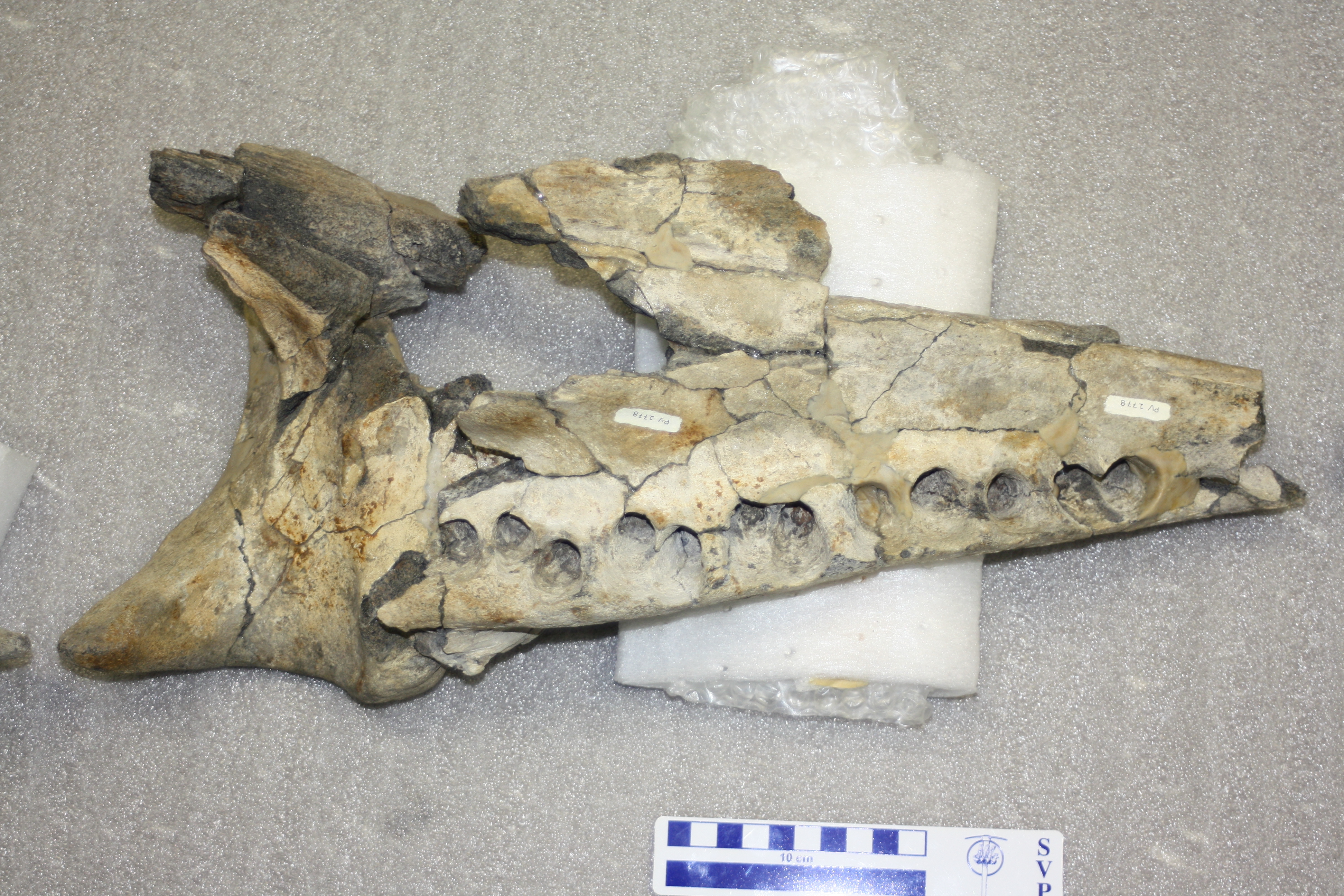
 The main skull part of ChM PV 2778, the holotype of
Coronodon newtonorum - as I first saw it in 2012.


 Placing the mandible of ChM PV 2778 (
Coronodon newtonorum) back into place for the first time.

In parallel, I had not looked at ChM PV 2778 - the "Newton" whale - for quite some time. I knew it had a partial mandible and much of a skull, and some earbones. I looked at it again in early spring 2022 and had the following revelations: 1) the mandible could actually be pieced back together, and it was in three pieces until I asked curator Matt Gibson to reconstruct it; 2) there was a nice isolated cheek tooth bearing the same number that at some point had been separated and was stored in a different cabinet. After the first revelation (which, mind you, was about the same time we realized that Al Sanders' conference abstracts indicated this specimen, and "Hoss" - ChM PV 5720 - had 12 instead of 11 mandibular teeth), I took a couple snapshots of the mandible fragments reassembled (not yet glued) and realized it looked quite a bit different from Coronodon havensteini. Instead of a straight ventral margin, the ventral margin in the "Newton whale" (ChM PV 2778) was convex. Likewise, the alveolar (dorsal) margin of the mandible was more strongly concave than in Coronodon havensteini. I looked again at the rostrum, and it too had a convex margin - indicating that ChM PV 2778 had a "smiling" profile of the lower jaw. Out of caution, we waited for Matt Gibson to glue it back together in case I was wrong - but it looked the same when I returned to take photos for the paper. This was the first time I thought that there might be a third species present - which surprised me: I assumed that we might have two chronospecies: C. havensteini in the late early Oligocene Ashley Formation and a younger chronospecies in the Chandler Bridge. I assumed this out of being cautious, conservative, and being a lumper. I swear to god, some folks - and dinosaur researchers in particular are guilty of this sin - seem to think every specimen is its own species and genus.

The holotype skull, teeth, and mandible of Coronodon newtonorum, ChM PV 2778, the Newton whale.

Upon further examination, I noted that the isolated cheek tooth of ChM PV 2778 is an upper tooth, and that it fit best into the sockets (alveolus) for the upper M1. The tooth is somewhat smaller than the M1 of Coronodon havensteini, an observation I duplicated for the lower 2nd premolar. The periotic is quite a bit more slender than in the holotypes of Coronodon havensteini or Coronodon planifrons - and in many respects, it resembles the juvenile specimens of Coronodon havensteini, despite itself being an adult specimen. One feature it shares with Coronodon planifrons is that long lateral tuberosity of the periotic - something lacking in Coronodon havensteini. As it happens, this specimen had a manuscript name and its own genus name - at this point, known only to those who visit Charleston Museum collections and look at the specimen labels where they're written - but the species name actually was proposed to be "newtoni" by Al Sanders and Larry Barnes. We went with this for two reasons: 1) it honored the earlier work by Al Sanders; 2) Claude and Albert Newton found and collected the specimen; and 3) the Newton family participated in 'volunteer night' at Charleston Museum for many years. There was just one catch, which we discovered during the proofing stage: "newtoni" honors only a single male member of the Newton family... and we didn't know which one, because Claude and Albert Newton both participated in the excavation. So, at the last minute, we changed the name to "newtonorum" to honor the whole family, and Claude and Albert in particular. I have not yet met anyone from the Newton family, but I'm hoping to now that the word is out! 

Line drawings of the skulls of Coronodon havensteini, Coronodon planifrons, and Coronodon newtonorum from the Oligocene rocks of Charleston, SC.

Other Specimens and the Importance of Publishing on the the "Dregs"

In addition to describing the new specimens of Coronodon havensteini and the two new species, I also wanted to take the opportunity to include a bunch of more fragmentary specimens. These are specimens that 1) could have conceivably been published as a separate paper but 2) the submission/revision process would have probably eaten up more time than the specimens are worth. Since my time is at a premium compared to earlier in my career, I decided instead to include these as 'accessory' data, so to speak. These come in two categories: 1) a bunch of isolated teeth and 2) a partial cranium from the Cooper River.

Isolated teeth of Coronodon from the Charleston embayment. Most of these are isolated, except for C-F, which are two associated teeth. Specimen K-L is one of the oldest discoveries made of Coronodon, found in 1974.

The isolated teeth constitute every single specimen we are aware of in museum collections at CCNHM and Charleston Museum. There may be more teeth in the South Carolina State Museum, which we did not sample. The lion's share of these lack good data and are identified as Coronodon sp., since it is unclear what species they belong to. One tooth, mentioned above, was identified as the distinctive M3 of Coronodon planifrons. Confusingly, this specimen was found at the same time and locality as the Coronodon planifrons holotype - but is from the same side as the M3, rather than the opposite side - so must represent a different specimen. The preservation is also not terribly similar. Altogether, there are 13 isolated teeth, found as early as 1973 (and possibly earlier). In contrast, I see at least a couple dozen teeth of the giant dolphin Ankylorhiza collected from the Charleston area and posted onto fossil groups on Facebook every year. I've estimated in the past that there are probably in excess of 100 Ankylorhiza teeth for every isolated Coronodon tooth. I'm only aware of three such teeth found since I started in 2015, and about 2-3 additional teeth remaining in private collections, in addition to the 13 isolated teeth we described. Coronodon is a very rare fossil, and I'm confident we probably have published virtually all of those discovered in the Charleston area.

 Partial skull CCNHM 8745 - a partial braincase indicating the presence of a third member of the Coronodon clade, now known as the family Coronodonidae.

A partial skull consisting of a phosphatized braincase with a bit of attached matrix has been floating around in our collection for years - sadly, the collector data is lacking, and the most likely collector - Stephen Miller - passed away a few years ago. According to Mark Havenstein, the preservation resembled fossils from the bottom of the Cooper and Edisto rivers, but not from the Wando River where the holotype of Coronodon havensteini and Inermorostrum xenops were discovered - river fossils there were typically grey to black, rather than brown. A clue to the origin of this specimen came from a sea turtle beak published in 2017 by Rob Weems and Mace Brown, who at the time, noted that the specimen had been collected from the Cooper River near Goose Creek, South Carolina. They identified it as the sea turtle Euclastes, and noted that fossils of the archaeocete Dorudon serratus were found with it on the river bottom and attributed to uppermost Eocene strata. The supposed archaeocete specimens, I was told on a different occasion, were this skull and an atlas vertebra thought to be associated with it - however, the atlas was actually from the giant dolphin Ankylorhiza. The Ankylorhiza atlas suggested Oligocene age - and indeed, there are not really any Eocene rocks in the subsurface there - not as shallow as the river bottom - and the river erodes instead into the Ashley Formation. While it's possible the specimens could have been transported from upstream, the vertebra is clearly from a large odontocete and the skull was a good match for Coronodon or something similar. Further, adhering matrix most closely resembles the Ashley Formation and there are these distinctive little convex clams preserved that are common in Ashley Formation nodules from Lowcountry rivers. I used to think that perhaps it was intermediate in morphology between Coronodon and Basilosauridae, but the more I looked at it the more it resembled Coronodon. The differences are mostly around the interorbital region of the skull: it has tiny, short nasal bones that are flat. Some aspects resemble Coronodon more than Basilosauridae and indicate this specimen belongs to Neoceti - the premaxilla contacts the frontal bone (in Basilosauridae the premaxilla terminates further anteriorly and is 'pinched' out by the nasal and maxilla), and the nasals are quite short with the 'blowhole' positioned far posteriorly, just somewhat in front of the eye sockets (resembling Coronodon) rather than halfway out on the rostrum (like Basilosauridae).

So, why publish on the "dregs" of collections? Two major reasons. First, out of scientific thoroughness: I've seen the dangers of "headhunting" - focusing on only the very best specimens, relegating the "dregs" of museum collections to oblivion. This is an underappreciated problem - from my perspective, I've seen so many specimens forgotten by disinterested or unthorough researchers sit untouched in museum collections. Why should we bother keeping such specimens if they're not going to be useful? I have the radical idea that *most* specimens should be published, as many, if not most, are more informative than is generally appreciated. Second, such specimens can provide unexpected benefits. For example: our suite of teeth sugests that Coronodon is quite rare as far as Oligocene marine mammal fosils are concerned. And, more critically, the 'ugly duckling' skull CCNHM 8745 may not be the most informative specimen of our study, but it does indicate that there are three coronodonid genera present in Oligocene rocks here: Coronodon, the larger Basilosaurus-sized cousin "Hoss" (e.g. ChM PV 5720), and the taxon represented by CCNHM 8745.

Introducing the New Family Coronodonidae 

The recognition of at least one other unnamed genus of Coronodon-like whale in this paper - along with a third containing the unstudied specimen ChM PV 5720, aka "Hoss" - led us to name the family level clade containing Coronodon. Unfortunately, Coronodontidae is preoccupied by some Devonian sharks, the type genus of which is Coronodus. Family names in taxonomy are pretty rigid: they have to have "-idae" onto the end of the type genus name. So, the other family I'm familiar with, Eomysticetidae? "-idae" is swapped for the last syllable in Eomysticetus. The correct familial emendation of Coronodon would be Coronodontidae - which is preoccupied. So, in lieu of that, according to the International Code of Zoological Nomenclature (ICZN), you just add "-idae" onto the end of the genus name, however it is spelled. Hence, Coronodonidae - which admittedly does not slip off the tongue quite as nicely as Coronodontidae, but there's nothing I can really do about it. Whenever "Hoss" gets described, it will be named within the Coronodonidae - and is already recognized as one in our paper. This family, by the way, is diagnosed by a long list of features including a blade-like part of the premaxilla that extends in front of the incisors, dorsally curving nasal bones, a lightly articulated (and possibly flexible) rostrum, deep zygomatic processes with enormous sternomastoid fossae just behind them, large cheek teeth bearing at least five mesial cusps, and cheek teeth that are highly emergent from their sockets - among a bunch of other features.

Next up: part 3 - new observations of the growth and functional anatomy of Coronodon and the surprising results of our phylogenetic analysis: is Coronodon actually a mysticete or could it be a late-surviving archaeocete?

Saturday, April 15, 2023

New fossils of Coronodon 1: new specimens of Coronodon havensteini

This Friday sees the publication of an expansive new monograph on the toothed baleen whale Coronodon havensteini in the journal PeerJ. I wish I could say I spent years on this, but as it happens, I wrote most of this in a marathon last summer to get it ready for an edited volume which was due in late September - in a stroke of irony, I assumed a monograph length paper was ok [narrator: it wasn not]. So, we formatted and submitted instead a couple weeks later to PeerJ. I had done some of the descriptive work on this in spring 2022, and much of the photography as early as 2017, but by and large, I cranked this monograph out in about five months. The monograph reports new specimens of Coronodon havensteini, names two new species - Coronodon newtonorum and Coronodon planifrons - and investigates the skeletal anatomy, body size, vertebral count, locomotion, biochronology/evolution, and phylogenetic relationships of Coronodon. The primary goal of this paper was to publish ALL known specimens of Coronodon. Future work coming down the pipe will include 1) a broader analysis of the dentition and feeding ecology of Coronodon and other toothed mysticetes and 2) a description of "Hoss", Coronodon's larger cousin.

For the second post in this series, click here.

For the third post in this series, click here.

Spoiler alert: this new study does NOT report much in the way of new evidence weighing in on the dental filtration hypothesis. There is another manuscript focused completely on that subject still in the works.

 Title page of our shiny new, enormous paper!

Introduction

Taking a step back - modern baleen whales are quite distinctive in lacking teeth as adults, and instead have the unique soft tissue structure that gives them their name - baleen - which is a gingival tissue growing from the palate and constructed out of keratin, the same material that makes up your fingernails, hair, and various other structures in mammals. Fossils of the earliest baleen whales, however, still had teeth - while this sounds surprising to most at first thought, it shouldn't be - having teeth is the norm in mammals, and of course dolphins (Odontoceti) have teeth - so of course at some point they had to have been lost. The first toothed mysticete fossils were not found until the 1960s, but generally recognized as archaeocetes until the 1990s. This is partly due to 1) overemphasis of study on Neogene (Miocene and Pliocene) fossil baleen whales which are very similar to extant species resulting in a poor understanding of what features evolved early at the mysticete-odontocete split and 2) the fact that most toothed mysticetes are quite similar to ancestral archaeocetes.

ChM PV 9163, Coronodon sp., likely the historically earliest Coronodon specimen ever collected, found in 1974.


 ChM PV 2778, the specimen that is now published as the holotype of
Coronodon newtonorum - the earliest discovered skull of Coronodon.

In the 1970s and 1980s a few low key discoveries were made here in Charleston. I say low key because for a long time they were never published upon - just presented at a few paleontology conferences in the 1990s, and colloquially known about within paleocetological circles for decades. Some archaeocete-like teeth were found as early as 1974. In 1978, a partial skull with teeth resembling basilosaurid whales was found in North Charleston near the Air Force base and what is now the Boeing plant, in what used to be a trailer park - a low income neighborhood that was re-zoned for an access road to a new Boeing plant, closed down, and razed (there's still nothing built there, by the way, it's just a buffer zone apparently). This specimen was collected for Charleston Museum and numbered ChM PV 2778. It came from a rock unit that would be named the Chandler Bridge Formation in 1986. ChM PV 2778 preserved a partial rostrum and a nearly complete mandible, generally revealing a basilosaurid-like whale with a skull about a meter in length. In 1989, a small skull, ChM PV 4745, was found revealing much more of the skull - a delicately constructed rostrum that is triangular and dorsoventrally flattened into the familiar shape we see in modern baleen whales - though the snout is a bit more robust than what we see in modern species, likely to accommodate some bite force during feeding. This specimen is somewhat older and was collected from the Ashley Formation. A third specimen, ChM PV 5720, indicated the presence of a Basilosaurus-sized whale - at least in terms of skull size - with even larger teeth, a slightly deeper and longer rostrum, with enormous temporal fossae housing massive jaw closing muscles. Charleston Museum curator Al Sanders teamed up with prominent marine mammal paleontologist (and Los Angeles NHM vertebrate paleontology curator) Larry Barnes* to study these fossils. They presented their findings at a couple of conferences in the mid 1990s, and never published anything on them; Al led the projects describing the toothless mysticetes from the Oligocene 'rocks' of Charleston which would eventually be published in 2002 as Eomysticetus whitmorei and Micromysticetus rothauseni. The fossils were by the early 2000s known colloquially as "archaeomysticetes". In 2003, Jonathan Geisler published the main study from his dissertation titled "Morphological Evidence for the Phylogeny of Cetacea", coauthored with Al Sanders, and reporting the first truly modern morphological phylogenetic analysis of Neoceti. This study included a bunch of as-yet unnamed odontocetes as individual taxa represented just by their specimen numbers - and also including two 'archaeomysticetes', ChM PV 4745 and 5720. Dozens of papers reporting fossil mysticetes used these codings, and Al was happy enough to host visitors at Charleston Museum to come see the specimens - with the understanding that the specimens could be coded, photographed, and examined, but not described or have published photos since the project was unpublished.

*Barnes is an imposing figure in our field - arguably marine mammal paleontologist Remington Kellogg's heir in North America - he dominated marine mammal paleontology until he retired from LACM about a decade ago and named in excess of 50 species.


My Ph.D. adviser, R. Ewan Fordyce, examining the rostrum of ChM PV 4745 in October 2012 during my east coast museum trip during the first year of my doctoral program.


The rostrum and braincase of ChM PV 4745 jigged up into near correct articulation at Charleston Museum in Oct. 2012.
 

Mace Brown taking a break from preparing specimens in what is now the CCNHM collections room, but what used to be a bit of a workshop and prep lab, with the "Wando whale", CCNHM 108, sitting on the table. October 2012.

Ewan Fordyce and Al Sanders talking about Oligocene cetaceans - two giants in marine mammal paleontology, College of Charleston, October 2012. Photo by Tatsuro Ando.

Eric Ekdale (left), Ewan Fordyce (center), and Tatsuro Ando (right) examining CCNHM 108, the Wando Whale, in October 2012 at CCNHM.


 Another great shot of Ewan and Al discussing 'archaeomysticetes' at CCNHM, October 2012.
 
 
The loose teeth of the "Wando whale", CCNHM 108, prior to having been glued into their sockets. CCNHM, October 2012.

 
The "Wando whale", CCNHM 108, as I first examined it in October 2012 at CCNHM. Just an utterly gorgeous specimen.
 

The lower left P3 of CCNHM 108, originally identified in our 2017 paper as the lower left P2, in my hand. CCNHM, October 2012.

I first met Al Sanders, and these specimens in October 2012, being one of those scientists who was given permission to examine unpublished specimens - which was quite generous of Al. Unbeknownst to us, Al had just retired from Charleston Museum. I had also just come from the 2012 Society of Vertebrate Paleontology conference in Raleigh, North Carolina, and Jonathan Geisler - who I had been working on some research with on the Pliocene whale Herpetocetus - told me about a neat collection under the care of someone named Mace Brown at the College of Charleston. He strongly suggested, in his often understated fashion, that I go visit the new College of Charleston Natural History Museum. I almost didn't, but Brian Beatty communicated a bit more directly with me and said "dude, it's incredible, don't even think of skipping it!". So, after my first full day at Charleston Museum, I took a very quick (and sweaty) walk down to the east side of Charleston, looked out at Fort Sumter, and then hustled back north towards College of Charleston - and found myself walking through this neat little campus with late 18th and early 19th century houses converted into academic departments, incredible oak trees, Spanish moss everywhere - and an incredible registry building that looked a *little* familiar (turns out it served as the magistrate building from the 2000 film The Patriot starring Mel Gibson). When I got to the museum, I immediately saw two incredible specimens: 1) a nearly complete eomysticetid! and 2) a nearly complete 'archaeomysticete' skull that rather looked like ChM PV 4745, the much smaller of the two. This new specimen was later catalogued as CCNHM 108. It had identical looking teeth and a similar triangular rostrum, and I immediately suspected the two were likely the same species, and at least the same genus. More critically, it told me that ChM PV 4745 was almost certainly a juvenile. I had two more days in Charleston, collected a bunch of data and photographs for my thesis, and then went back to New Zealand and focused on eomysticetids and early mysticete phylogeny - with these specimens really leaving a serious impression: all prior studies really left out some big gaps in our knowledge of early mysticete evolution. The 'archaeomysticetes' really needed to be published! On my second day of visiting CCNHM, I sat in the Five Guys on King Street wolfing down a burger and thought "huh, this would be a nice place to work."


The holotype mandible of Llanocetus denticrenatus on display in the Sant Ocean Hall at the Smithsonian (USNM) and the published figure from Mitchell (1989).


A cast of what eventually turned out to be the holotype skull of Llanocetus denticrenatus, on display in the Sant Ocean Hall at USNM. As of April 2023, the actual skull remains at the University of Otago, but will be shipped back to USNM in the future. Originally given an Otago catalog number until it was discovered to represent the same specimen as the holotype, after which an agreement was reached between the two institutions.

A discovery made in parallel - Llanocetus denticrenatus - was originally named in 1989 by Ed Mitchell - a strange toothed mysticete known only from a few fragments and an endocast from the uppermost Eocene La Meseta Formation of Seymour Island, Antarctica. Mitchell's paper had surprisingly far-reaching conclusions in the paper about early mysticete evolution. As it happens, the original fragments had been collected in the 1970s and deposited into USNM (Smithsonian NHM) collections. My Ph.D. advisor, R. Ewan Fordyce, had discovered a nearly complete skull in 1986 at the same locality, and had most of it prepared when Mitchell visited in the late 1980s. Eventually, it turned out that the skull collected and prepared at University of Otago was actually the very same specimen that Mitchell (1989) published on and fragments collected in 1986 matched up with those collected in the 1970s. Fordyce was surprised to see the fragments published in the 1989 article, and the 'scooping' is partly why the rest of the skull was not formally published until 2018.


 The holotype skull of the small aetiocetid whale "Chonecetus" goedertorum, now Fucaia goedertorum, from Barnes et al. (1995). Species discovered by and named after Jim and Gail Goedert; collected from Twin River Quarry in the Pysht Formation of Washington, USA.

Since the 'archaeomysticetes' were first informally announced, and the original scraps of Llanocetus published, many discoveries have been made in parallel and the field has had to grow and move without these fossils, leaving them behind on the wayside, so to speak. In 1995, a landmark paper by Larry Barnes, Masaichi Kimura, Hitoshi Furusawa, and Hiroshi Sawamura reported a number of new fossils to the Aetiocetidae. Previously, the family was only known from Aetiocetus cotylalveus and Chonecetus sookensis - the former was collected by west coast hero Doug Emlong, and published by him in his only paper in 1966 - and the latter was named by Loris Russell two years later based on an "indifferently preserved" braincase, to quote Ewan Fordyce. Both Chonecetus and Aetiocetus were considered archaeocetes, though Russell noted some odontocete-like features in Chonecetus.This new study by Barnes et al. named the new species Chonecetus goedertorum based on a small, narrow-snouted skull from the Pysht Formation of Washington, USA; the new genus and species Ashorocetus eguchii based on fragmentary Chonecetus-like skull from Japan; Morawanacetus eguchii, based on a somewhat more complete braincase and a large multicuspate molar; and three new species of Aetiocetus: Aetiocetus weltoni from the Yaquina Formation of Oregon, Aetiocetus tomitai from Japan, and the more complete Aetiocetus polydentatus, also from Japan. This study established that 1) aetiocetids were actually toothed mysticetes and 2) toothed mysticetes, at least in the North Pacific, were very diverse. In 2006, Erich Fitzgerald published a spectacular fossil from his Ph.D. research - the new short-snouted, big-eyed toothed mysticete Janjucetus hunderi from the Oligocene of Australia - which indicated that some toothed mysticetes were adapted towards macrophagy, similar to archaeocete whales. This was followed in 2010 by a redescription of Mammalodon colliveri, originally named in the 1930s from a nearby locality, which turned out to be closely related to Janjucetus as well as a benthic suction feeder. 

 Lateral palatal foramina and sulci in a modern mysticete (minke whale, A-B) and in the toothed mysticete Aetiocetus weltoni (D-E), from et al. (2008). This is now perhaps the single most controversial figure in the history of the study of baleen whales.

The most controversial study, however, was published by San Diego Natural History Museum curator Tom and colleagues in 2008, reporting lateral palatal foramina - the osteological correlates of baleen - in Aetiocetus weltoni. They proposed that aetiocetids had teeth and baleen at the same time, and highlighted a stepwise hypothesis for the teeth to baleen transition: toothed ancestry, followed by some species with teeth and baleen, followed by the loss of teeth. Tom showed me the palate of Aetiocetus weltoni in 2007, after I had seen his presentation at the 2006 Society of Vertebrate Paleontology conference and been skeptical - but when I saw the skull in person, I thought "holy shit, he's right!". After the paper came out in 2008, I thought "great! What's next for mysticete paleontology?" and had no clue that we'd still be fighting about it fifteen &%*-ing years later. It's literally been fifteen years. Some studies have tried to argue that baleen wouldn't work with interdigitating teeth; that the baleen itself is not preserved; that the tooth wear indicates suction rather than filter feeding; that maybe the homology is wrong; that hippos have similar structures in their palate yet lack baleen (they're not homologous in hippos); and that mysticetes actually lost their teeth before they gained baleen. All of these are tough sells (in my opinion) and several have been appropriately discounted in the literature (and others are on their way into the rubbish skip at the moment - stay tuned). All of these advances were made without anyone really being able to talk about the 'archaeomysticetes' or Llanocetus in detail.

 Figure 1 from the original paper reporting Coronodon havensteini (CCNHM 108), Geisler et al. (2017).

Coronodon havensteini - what we originally reported

Shortly before my Ph.D. program concluded I was invited by Jonathan Geisler and Brian Beatty to help them wrap up study of CCNHM 108 - the beautiful skull I had seen at CofC in 2012 that I thought was the same taxon as ChM PV 4745. The manuscript focused only on this specimen - and since I had by this point locked down a job at CofC, I'd be able to directly record more observations, measurements, and photographs of the specimen in person. We worked on this for my first two years at CofC, and published "The Origin of Filter Feeding in Whales" in June 2017. In our paper, we named CCNHM 108 as the holotype specimen of Coronodon havensteini, finally getting a name attached to the 'archaeomysticetes'. We noted that in addition to an overall morphology that looks like a basilosaurid at first glance, including basilosaurid-like teeth - there are a number of unusual points of feeding morphology that suggest something other than basilosaurid-like feeding.


 The mandibular dentition and interdental slots of Coronodon havensteini and a diagram showing water flow through the closed mouth of Coronodon - mandible of the basilosaurid Zygorhiza for comparison. Also tooth erosion and comparison with a Mesoplodon tusk. From Geisler et al. (2017).

First and foremost, Coronodon at first glance appears to have heterodont teeth - incisors, canine, premolars, and molars - which is to be expected for an early toothed mysticete. However, it has actually evolved towards having cheek teeth that are more uniform than in basilosaurids - the premolars and all the molars are similar in size and cusp number, and cannot be easily identified to position without 1) process of elimination and 2) having teeth with complete roots and nice sockets in the mandible. In a way, the postcanine teeth have evolved a degree of homodonty - but not the way you normally think of it in the context of homodont teeth in dolphins, where they're all conical. These cheek teeth, at least in the mandible, also overlap like roof shingles: the posterior half of each tooth is slanted posterolaterally, and each tooth overlaps the next one by nearly a centimeter. We proposed that this could act as a dental filter, as the interdental slots formed by these overlapping teeth are quite narrow. Tooth wear further supported this idea, as the only appreciable apical tooth wear was on the tips of the anterior teeth - incisors through the second premolar. Aside from one or two broken cusps, the cheek teeth are devoid of tooth wear. The wear facet made by the upper and lower teeth locking together are also completely on the side of the tooth and on the root, rather than forming a slicing facet at the tip of the crown like their basilosaurid ancestors - which is the same way we see how dog and cat carnassials slice against each other. This suggests that Coronodon was preferring to bite with its conical anterior teeth and avoiding slicing and dicing (and chewing in general) with the posterior teeth. We also noted that the teeth are pretty emergent from their sockets - e.g., they stick out a LOT - perhaps to help enhance the dental filter. Additionally, the rostrum (snout) of Coronodon is quite wide, like in toothless mysticetes, and quite unlike most other toothless mysticetes, it has reduced bony connections between the bones of the rostrum (more on that).

 Our partial cladogram and ancestral character state reconstruction of tooth spacing from our 2017 paper on Coronodon (Geisler et al., 2017).

We interpreted this as evidence that Coronodon was adapted to filter feeding with its teeth - and drew comparisons based on similar patterns of tooth wear with the leopard seal Hydrurga leptonyx, which filter feeds for krill much of the year and bites larger prey (e.g. penguins) the rest of the year. Hydrurga has tooth wear concentrated on the canines and incisors and the cheek teeth are relatively unworn. To further test this, we included Coronodon into a new matrix that was sort of a combination of Jonathan Geisler's original matrix from his dissertation and my thesis matrix - we primarily used the former but included a lot of new characters from my thesis. Because the skull of Llanocetus was still unpublished, we used codings from Erich Fitzgerald's paper on Mammalodon. We were interested in tracking the evolution of tooth spacing, so we recorded the relative distance between teeth divided by tooth diameter - Coronodon, with its overlapping teeth, had a negative number, whereas aetiocetids had gaps between their teeth at least 0.5 to 3 times the diameter of teeth (0.5-3). Llanocetus had a similar measurement. We generally found a trend towards wider and wider tooth spacing in later diverging toothed mysticetes - and suggested that perhaps Llanocetus, along with aetiocetids, may have had baleen. 

Analysis of tooth sharpness in early mysticetes by Hocking et al. (2017) - CCNHM 166 is now Coronodon planifrons. A fine study, but only if you take the assumption that teeth can only be used for filter feeding if they function the same way as in the extant seals Hydrurga and Lobodon - which was not the argument we made, we argued for the use of interdental slots between the teeth.

This admittedly provocative hypothesis was quickly criticized, and a few months later, our Australian colleagues, led by David Hocking, published an analysis of tooth sharpness and found that, indeed, teeth of Coronodon sp., CCNHM 166 (now Coronodon planifrons) - are sharp. To be precise, the notches between the cusps resemble what you see in the carnassial notch of extant big cats. Armed with this data, they published a paper with the bold title "Ancient whales did not filter with their teeth". Admirable, but it's based on a single aspect of the feeding morphology of Coronodon. We didn't even focus on the cusps- we focused on the interdental notches between the overlapping teeth (which, by the way, still exist). 

What's new with Coronodon havensteini?

I'll be covering the two new species in a followup blog post, but before that, let's cover the new specimens and their insights into the anatomy and growth of Coronodon havensteini. We report in this paper three new partial skulls of Coronodon havensteini: ChM PV 4745, the juvenile specimen originally under study by Al Sanders, CCNHM 8722, a second juvenile skull collected just in the past couple of years (nicknamed the Volcko juvenile, after the collector, Jeremmiah Volcko, who co-discovered the Coronodon planifrons, which for a long time was called the Volcko whale or Volcko Coronodon), and CCNHM 164, an old adult nicknamed the Coosaw whale or Coosaw Coronodon.


 The ontogenetic sequence now established for Coronodon havensteini - two young juveniles or calves, CCNHM 8722, ChM PV 4745 - the adult holotype CCNHM 108 - and the old adult, CCNHM 164.

The juvenile specimens are small - about 2/3 maximum size - so we're talking about large calves here. Not really babies, but individuals that are small enough to tell us about early growth. Both juveniles happen to have skulls with snouts (rostra) that are surprisingly the same proportions as the adult holotype - same length relative to the braincase, and same length to width ratio. I was not expecting this: in cetaceans, the rostrum is usually shorter in juveniles and lengthens later on during growth. Likewise, another surprising proportional change during growth is that the sagittal crest - a tall ridge of bone on the dorsal side of the braincase forming muscle attachment for the jaw closing muscle (temporalis) gets considerably longer from juvenile to adult. Juveniles tend to have bigger braincases than adults and they grow into them, so to speak, but still, juveniles also tend to exhibit archaic or primitive features that change into the more derived adult morphology during growth. A long sagittal crest is the plesiomorphic condition, yet this increases in length during growth! It's very possible that this ontogenetic sequence is inherited from basilosaurid whales - we know very little about their growth. The upper teeth in ChM PV 4745 are preserved in situ, and they are not very emergent from the upper jaw (maxilla) - in fact, the base of the enamel crown is still obscured by the bone in side view. This tells us that eruption of the teeth is delayed late into growth. Additionally, the teeth in the upper jaw overlap one another, just like the mandibular teeth of the holotype specimen. This is almost certainly a consequence of having to erupt enormous adult teeth into a small head - and I've seen baby and juvenile sea lion jaws where the teeth are overlapping early and and rotate during later growth and separate. These juveniles also have different looking earbones than the adult: the periotic/petrosal bones are narrower and more gracile, and the tympanic bulla is smaller.


Adult mandibles of Coronodon havensteini: holotype, CCNHM 108 (top) and old adult, CCNHM 164 (bottom).


 Upper teeth of
Coronodon havensteini - the three teeth up top were all we had in the holotype, but these somewhat more worn teeth of the old adult, CCNHM 164, help fill in some of the morphology.

The old adult specimen, CCNHM 164, is missing a rostrum but has a partial braincase that is quite fractured. It also has many more of the upper teeth preserved than in the holotype - and the teeth in general are a bit more worn down, as to be expected in an old adult. There are also many more incisors: these are surprisingly tiny, far smaller than the reconstructed teeth present in the holotype specimen - 1/2 to 2/3 the diameter, and with smaller crowns. This suggests that Coronodon had comically small grasping teeth in the front of the jaw in comparison to the rather large premolars and molars that overlap one another. CCNHM 164 also has a disarticulated rostrum, and we can see the articular surface on the premaxilla (the bone that makes up the middle of the snout, and houses the incisors; all other teeth are embedded in the maxilla). First, this means that the rostrum was loosely articulated enough that it was able to fall apart after death: the premaxilla fell off the skull, and the maxilla from the premaxilla - in an old adult. This means that the rostrum of Coronodon is lightly built and did not fuse at all during growth - in other words, this is not just another suture that will firm up during growth. The rostrum of Coronodon was also probably kinetic to some degree - an adaptation we see in modern baleen whales, thought to be correlated with dispersing the mechanical stresses involved with filter feeding, but admittedly poorly understood and understudied.


Ontogenetic sequence of Coronodon havensteini periotic bones. From top to bottom: juvenile, CCNHM 8722; juvenile, ChM PV 4745; adult holotype, CCNHM 108; old adult, CCNHM 164.

CCNHM 164 also has periotic bones that are really, really inflated: along with the gracile juvenile periotics, this indicates that the periotic becomes thicker and more robust during growth, without getting any longer. The tympanic bulla also grows from juvenile to adult - CCNHM 8722 has the smallest bulla, ChM PV 4745 is a bit larger, and CCNHM 108 has an even larger bulla (none preserved in CCNHM 164). This same pattern is known in basilosaurid whales as well as the eomysticetid whale Waharoa ruwhenua, but in modern baleen whales, so far as we can tell, the bulla is adult size at birth. The same is true for odontocetes which have been studied.

Postcranial bones of Coronodon havensteini - holotype specimen, CCNHM 108, on left, and referred old adult specimen, CCNHM 164, on right.


Composite skeletal reconstruction of Coronodon havensteini, using lumbocaudal series count and morphology from Coronodon planifrons. Terminal caudals, shape of most of the neural spines, forelimb (partial scapula only), hyoids, and hindlimb unknown.

CCNHM 164 also has quite a bit of the vertebral column preserved, including three lumbar vertebrae and nine thoracics; in concert with the nearly complete lumbar and caudal series of Coronodon planifrons, we now know that Coronodon likely had seven cervical vertebrae, nine thoracic vertebrae, ten lumbar vertebrae, and at least 20 caudal vertebrae. The vertebrae are short and differ from the 'coke can' shaped vertebrae in Basilosaurus - but closely resemble the vertebrae of Dorudon atrox in many respects. The posteriormost caudal vertebrae of Coronodon planifrons are rectangular, which means that Coronodon had caudal flukes - however, already known for Basilosauridae. In contrast, the caudals are all relatively wide - and so Coronodon did not have a narrowed caudal peduncle as in modern cetaceans. I proposed that a narrow peduncle (tail stock) was absent in Xenorophidae back in 2017 - and confirmed this when we reported on Ankylorhiza - and now doubly confirmed in an early mysticete. The largest vertebrae - the ones that are the longest and widest - are in the posterior lumbars and anterior caudals - indicate that Coronodon did have a slightly stiffened peduncle, but there are not clear regions of the vertebral column with different proportions like we see in odontocetes, which places Coronodon into the "Pattern 1" swimmers - "Pattern 1" swimmers, according to Emily Buchholtz, swim by undulating their entire vertebral column. "Pattern 2" swimmers have some stabilized parts of the vertebral column and others that are more flexible, and "Pattern 3" swimmers have short regions where most of the dorsoventral undulation (bending) happens - almost like designating a short section of vertebrae like an 'elbow'. Lastly, when you add up all of the vertebral lengths, skull length, and reconstruct the additional length occupied by the cartilaginous intervertebral disks - we get a body length of about 4.8-5 meters. This is pretty short - and indicates that Coronodon had a comically large head. Which, if you think about it - is actually a baleen whale feature. Basilosaurids have relatively small noggins, and some modern baleen whales have skulls that are about 1/4 to 1/3 of the entire animal's length - with the most titanic heads belonging to the right whales. (Absolutely larger skulls are in the blue whale, though they are proportionally much smaller). What's great about fossils like these are that we can, even with a somewhat incomplete vertebral column, evaluate methods used to estimate body length. We tested the skull width body length estimates published by Pyenson and Sponberg (2011) and found that their equations underestimated the body length of Coronodon by about 12-15% - not great. I actually pointed this out in my papers on Ankylorhiza and Tokarahia, with even more extreme underestimates - it seems these equations are not well-adjusted for the skeletons of the earliest Neoceti and routinely underestimate their body length.


 The holotype skull of Coronodon havensteini (CCNHM 108): A, with only the original teeth; B, after cast teeth were installed and reconstructed parts of the skull painted to match; C, skull and mandible in approximate occlusion. This really shows that the anterior tip of the mandible, including the entire alveolus for the first incisor, is missing.

Tooth Count in Coronodon and Neoceti

When we published on our paper in 2017, we focused exclusively on CCNHM 108 and deliberately had blinders on - we did not want to include any observations on specimens still under study by Al Sanders. We interpreted Coronodon havensteini as possessing 11 upper and 11 lower teeth - and thought that the mandible was complete. In the early stages of this monographic work, I remember being troubled by the posteriormost teeth - in order to make the teeth in the front of the mouth interlock, there would be a 3-4 cm gap between the mandible and the jaw joint on the skull! Also, the coronoid process of the mandible abutted the back of the eye socket and prevented complete closure. What was the deal?

Revised tooth count in Coronodon havensteini: top, showing interpretation of only 11 mandibular teeth, counting from the back of the toothrow, from Geisler et al. (2017), and bottom, updated counting, based on articulation of skull and mandible and new information from other specimens showing a count of 12 mandibular teeth, and indicating that the mandible of CCNHM 108 is missing the lower first incisor alveolus.

Unfortunately, we did make a bit of a bonehead mistake: In a couple of conference abstracts from the 1990s, Sanders and Barnes had actually written that two of their specimens - ChM PV 5720 and ChM PV 2778 - had twelve mandibular teeth. I went over to Charleston Museum, inspected both specimens, and confirmed this observation. Damage to the tips of the mandibles in CCNHM 108 had removed the socket for the lower first incisor! This meant that our lower third molar (m3) was actually the fourth molar (m4). When the mandibular teeth are placed into correct occlusion, the jaw articulates with the skull properly, and the last mandibular tooth sits just posterior to the posteriormost upper tooth - like basilosaurids, which have one fewer upper than lower teeth, the last mandibular molar (m3) sits posterior to the last upper molar (M2). These findings indicate that Coronodon is actually polydont: polydonty is the evolution of a higher tooth count than the typically constrained count of 11 teeth per quadrant. Only a few cetaceans have evolved polydonty: a few toothed mysticetes (e.g. Aetiocetus, Mammalodon), odontocetes, modern mysticetes (the fetal dentition is polydont), the giant armadillo Priodontes, and manatees (sort of; they clone the last molar and just keep making them, so their dentition is sort of immortal). There are a number of other toothed mysticetes that very clearly are not polydont - Janjucetus, Fucaia, probably (but uncertainly) Llanocetus - so it is unclear if polydonty evolved once or twice. Likewise, there are some early odontocetes that are very clearly non-polydont as well, including Simocetus - and others that are quite polydont, including the Xenorophidae, Waipatia, and Ankylorhiza, that have in excess of 13 teeth per quadrant. If it evolved once at the base of Neoceti, then a bunch of lineages immediately lost it. We don't necessarily understand the genetic underpinnings of polydonty, so it's possible that whatever genetic underpinning permitted polydony evolved only once, followed by increases and decreases in tooth count (with the toothless suction feeder Inermorostrum representing extreme tooth count reduction!). Indeed, Coronodon demonstrates that polydonty is probably ancestral to all Neoceti (odontocetes + mysticetes).

Next up: part 2, introducing the two new species; and later on: part 3, broader implications for our knowledge of early mysticete evolution and their phylogeny.

References

, , , . . . The Island Arc 3(4):392-431.

, . . . Journal of Vertebrate Paleontology 16:21A.

, , , . . . Systematic Biology 57:15-37.

, . . . Current Biology 28:1670-1676.

, , , . . . Current Biology 27(13):2036-2042.

, , , . . . Biology Letters 13:20170348.

. . . Canadian Journal of Earth Sciences 46:2219-2235.