Monday, December 14, 2009

Partial mysticete jaw (more fossil preparation)

About two years ago I came across a partial lower jaw of a small baleen whale in a cliff. The outwardly bowed part of the jaw was facing the outside of the cliff, and the middle had eroded away, leaving the anterior and posterior ends. I immediately collected the posterior end, which was (and currently still is) in a small nodule, and awaits further preparation. However, what is preserved indicates it belongs to Herpetocetus - my favorite cetacean. The posterior mandible of Herpetocetus is very distinctive; the three main features of this part of the jaw (the coronoid process, mandibular condyle, and angular process) are all anteroposteriorly elongate relative to other mysticetes, with the angular process projecting posteriorly as a flat flange. Anyway, as the specimen isn't prepared yet, I'm not going to show you pictures of the interesting part.

After I collected the posterior part, I tried to excavate the anterior portion; I successfully removed two segments which broke along natural cracks, and a third portion (which I figured at the time was the anterior tip) which was stuck in a nodule, and was rather stubborn. I decided to leave that part in the cliff, and return later under more favorable conditions. The next few visits it remained, and I checked up on it; I figured noone else would disturb it (or even spot it). Well, I got lazy, and over thanksgiving, I returned with the intention of collecting it, and some fresh pick marks were around it, and whoever it was had chipped away a little bit of bone, much to my chagrin. Anyway, I excavated the rest of it immediately; fortuitously the other person had started a trench around the concretion, so it took a mere 5 minutes to carve around it and pop it out - and it did make a 'pop' sound and land with a resounding thud on the beach sand, to which I said to myself "if I had known it would have been that friggin easy, I woulda done it two years ago!".The anterior dentary before and after chiseling.
The problem was, I had no idea whether or not the two pieces would even connect, given the damage done by the other collector, and two years of erosion. And I had to fly with the fossil in my duffel bag all the way back here to Montana to find out. I "gingerly" chipped away some pieces of the concretion with a rock hammer and chisel, which split the rock from the bone perfectly.

The anterior 1/3 of the Herpetocetus mandible.

Then, when I got back to Bozeman, I took the fossil to campus and nervously matched up the two sides - a lot of bone was definitely missing, but (thank god) the two pieces matched up along the ventral side of the bone, preserving the natural length (which is kind of moot anyway, given that I'm missing the middle - kind of, because I measured the missing distance when I was in the field). Some major acid preparation is in order, as well as some serios airscribing and microblasting on all three pieces. These specimens are very similar to mandibles of Piscobalaena, a cetotheriid from the Pliocene of Peru, and the probable sister taxon to Herpetocetus (Bouetel and Muizon, 2006). To be totally honest, the piece of the mandible I've shown you looks pretty damn similar across much of Chaomysticeti (baleen bearing mysticetes), with the possible exception of balaenids.


The mandible of Piscobalaena nana from the Early Pliocene of Peru (from Bouetel and Muizon, 2006)

This is one of five partial Herpetocetus dentaries I've collected from the Purisima Formation (none are currently in museum collections at UCMP or SCMNH), and is the second most complete specimen; one is a complete, ~4' long, very large and robust dentary, and the others are all posterior dentaries (i.e. posterior 1/3). Two of these are extremely small (i.e. one is a fragment where the shaft of the dentary was only 2.5cm high), and likely represent neonates or extremely young individuals. These specimens, along with a nearly complete skull, a partial skull, half a dozen petrosals, and several tympanics will be the subject of a study by Jonathan Geisler and myself. In addition, there are two more possible (one definite) Herpetocetus crania in-situ, which will (hopefully) be excavated over winter break.

For more information on cetotheriids, see Alton Dooley's recent post on cetotheriids at Updates from the Vertebrate Paleontology Lab.

Bouetel, V., and C. de Muizon. 2006. The anatomy and relationships of Piscobalaena nana (Cetacea, Mysticeti) a Cetotheriidae s.s. from the early Pliocene of Peru. Geodiversitas 28:319-395.

Sunday, December 6, 2009

Fossil preparation - odontocete tympanic

About two or three summers ago I collected a beautiful little odontocete tympanic from the Purisima. Problem was, I only found out it was beautiful (past tense) after it sat in about twenty or thirty pieces. Because the part that was exposed looked like some other type of bone (and not an odontocete tympanic) I mis-estimated how sturdy the fossil was, and it exploded as I carved matrix away from it. I have since not repeated the mistake. Anyway, the fossil has sat in pieces in a plastic bag for two years, and I finally got the courage to glue it back together. I say courage because 1) I was somewhat embarassed by this damage, and 2) I was nervous to piece together all these tiny fragments. Below is a photo of what I had to work with.
Tympanic fragments prior to preparation.

I began by finding the pieces of the robust involucrum, which is the thick portion of the cetacean tympanic. There are more or less three major portions of the tympanic: the involucrum (frequently the only preserved part), the posterior process, which attaches to the posterior involucrum, and the paper-thin involucrum (which in odontocetes is usually under 1-1.5mm thick, hence the overall fragility of these elements). Then, I started finding matching pieces, and gluing these to the involucrum.

The tympanic after 30 minutes of preparation; some of the outer lip fragments have been glued in place. Lateral view (top photo) and anterior view (bottom photo) - note the matrix-free tympanic cavity.

After a couple more hours, I was able to finish gluing back most of the outer lip of the tympanic, as well as the posterior process.

Tympanic before (left) and after (right) addition of the sigmoid and posterior process.

Tympanic in dorsal (left) and lateral (right) aspects.

After the fossil was glued together, it became very obvious that this was a tympanic from the "river dolphin" Parapontoporia wilsoni, which has a small posterior process, a sharp anterior apex of the bulla, and most characteristically a laterally inflated outer lip, not seen in any other Purisima odontocete (for which tympanics are known, and out of the given tympanic sample from the Purisima Fm.). As far as crania, jaws, periotics, tympanics, and parts thereof go, Parapontoporia is by far the most common Purisima odontocete (i.e. between collections at UCMP, SCMNH, and LACM go, there are roughly a dozen nearly complete crania known, rostrum not included).

Newly prepared tympanic (right) side by side with another very well preserved Parapontoporia wilsoni tympanic.

All in all, I was extremely pleased; in one afternoon I had turned a pile of fragments (which I had virtually no hope for) into a beautiful little specimen. All but three tiny fragments under 5mm in size were glued on; the other ones probably attached to the margin of the outer lip, which may require fragments lost during collection (or, conversely, pieces pulverized). But let's not split hairs here - this by far was the most damage I've ever done to an odontocete tympanic, to the point where I was embarassed to even think about it; and now, it's one of the nicest I have. The positive side to this inadvertently destructive mode of
collection was that all the matrix was absent from the tympanic cavity, unlike the specimen on the left in the above photo (where the matrix inside was actually phosphatized, but phosphatic 'cementation' had fortuitously not formed an overgrowth around the rest of the bulla - best case scenario!). This is one of about a dozen and a half or so odontocete bullae I've recovered from the Purisima.

Petition to save the UBC track site in New Jersey

An early Jurassic dinosaur track site in New Jersey is currently in danger of being destroyed by development for a new set of high-end condos. The track site is directly adjacent to a park, and if the park boundary is extended only 200 feet, the locality can be preserved for posterity (and I believe *most* or *some* of the condo development can continue as well, so that would be more or less a win-win for both sides).

While many folks don't believe in online petitions, the "Help Save Capitola!" petition catalyzed opposition in 2003 and 2004 to help defeat the seawall that would have been built along the Capitola cliffs; that locality was saved, due in part to the petition I drafted during my freshmen year of college.

Anyway, the petition can be viewed here and signed here. I highly suggest anyone worth their salt as a paleontologist or fossil enthusiast to sign the petition; it broke 1000 signatures friday morning, and as of now, has 1804 signatures.

Saturday, November 21, 2009

Away For Thanksgiving

Sorry about the lack of posts over the last week or so; in a few hours I will be flying to California for Thanksgiving break, and this time will have 8 days (and thus, another 10 days before I write on here again) - my longest Thanksgiving ever, I think. What's this mean for me? Well, obviously I'm going to eat a lot of turkey with some of my relatives (my favorite set of cousins will be there; awesome). However, Thanksgiving is the first iteration of my 'real' field season.


Every spring, all my friends get antsy and extremely excited for the summer field season with Museum of the Rockies; they spend about two or more months digging up dinosaurs in the badlands of Montana - usually the Judith River Formation and the Hell Creek Formation, and formerly the Two Medicine Formation (although a few camps have been there within the last few years).

Summer has a completely different meaning for me; there is very little erosion on the coast, and summer is typically a season of crowded beaches, traffic over highway 92 and 17, high school kids are out of class and go smoke pot on the beach (and usually sit right on the outcrop, and are too dumb to move). Beach sands typically accumulate during the summer, piling up ever higher, foiling any attempt at collecting material below the high tide line. The lack of rain and storm waves means that most exposures by now are coated with a healthy coat of dust and grime, rendering any possibility of spotting smaller shark teeth and bird bones improbable.

Winter, on the other hand, is my field season, albeit short. Winter storms clean the cliffs off, erode the cliffs, and strip sand off beaches, allowing collecting from the wave-cut platform and the base of the cliffs (sand movement during storm and fairweather usually abrades the portions of cliffs that are buried during the summer as well). Additionally, the cold temperatures drive away the throngs of people who flock to the boardwalk and Santa Cruz beach during the summer, which means all I have to deal with is rush hour traffic.

Winter obviously has its drawbacks; 1), it's cold; 2), it's rainy; 3) the hours are short; and 4), the reduced sand level (sand gets stored in offshore sand bars during the winter months) often means access to certain areas is much trickier, often requiring some creativity and flexibility (i.e. lots of climbing, boulder hopping, etc.). This winter is an El Nino event, which excites the hell out of me - some vicious winter storms this year would be awesome. Last winter was fairly lame (although the little rain we did receive made it easy to get out), although I did pick up a nice delphinoid odontocete skull and a partial Herpetocetus dentary. Winter 2007-8 was just amazing, however; a nearly complete Carcharocles megalodon tooth (the day before christmas, no less!), only the second known for the Purisima (and only one available to scientists for research - will be donated to UCMP in the next few months), as well as a 40-50% complete female fur seal skeleton, a juvenile Herpetocetus dentary, and a smattering of pinniped bones.

I wish I really could properly convey my excitement to you - Thanksgiving field trips are the prelude to the really good 'stuff', so to speak, although storms typically haven't cleaned the cliffs and lowered sand levels enough to be terribly different from summertime. However, the three months between summer and thanksgiving makes it seem that much more prolific. I'm also uber excited, because I will be visiting some old localities I haven't been to in a while.

On a final note, for the last three and a half years I've been working on a manuscript version of my undergraduate research project, which was the description and analysis of a new, probable late Pliocene assemblage in the Purisima Formation. In 2007 I sent a copy of this to a couple colleagues of mine (Frank Perry, Santa Cruz Museum of NH, and Chuck Powell, Menlo Park USGS); unfortunately, my writing skills were relatively poor at the time, the manuscript was incomplete, figureless, and had older 'chunks' from previous drafts embedded, much like an accreted terrane that is then subjected to regional metamorphism; a really ugly, foul smelling, nasty thing that no one should bother looking at (can't you tell I enjoyed metamorphic petrology?!).

Needless to say, after reading the revisions, I felt pretty guilty that I had bothered having them proofread it in the first place. In any event, I've spent the last month seriously reconsidering every word in the manuscript, and rewriting it piece-by-piece into a coherent piece of literature, complete with submission-ready figures; last night, I emailed it out (again), and hopefully this time there will be less 'red'. I showed up at our local sports bar (Spectators, my third home; second home being my apartment, first home being my office) and let the 60-page paper drop to the bar table with a resounding 'thud', which, oddly enough, drew applause from my (drunk) friends and a high five from my friend (Hi Christina!) who's going to proofread that copy. Within seconds, a red pen was already out, which had crossed out my last name and replaced it with "boogernecker". Thanks, guys.

Anyway - Happy Thanksgiving!

Tuesday, November 10, 2009

New artwork - skull drawings

Well, folks, I've been a little busy recently, but in the last month or so I've gotten back into artwork in a big way; every once in a while I'll do a drawing I don't like, and I won't draw for a long time. This first drawing was formerly one that I got bored with a couple years ago. Then, after teaching a lecture on scientific illustration, I saw the bare bones shading I had done, and all of a sudden Dorudon started cackling and laughing at me; I took it as a challenge. I worked nonstop for about 10 hours, and finished it up at 3 in the morning that night. I started this thing in 2007.
Cranial drawing of the archaeocete whale Dorudon atrox,
from the Eocene of Egypt. 2d, graphite, 2007-2009.

The next drawing I completely forgot about; this is a drawing of a walrus (Odobenus rosmarus) cranium on display at the Smithsonian. This was shaded waaay too much, but the particular style of drawing I developed in high school called for really dramatic contrast and lights and darks, so that's what I'm stuck with.

Cranial drawing of modern walrus, Odobenus rosmarus. 2d, graphite, 2007.

The next drawing is actually pretty big; I finished this one on sunday. This is a lateral view of a 7-9' long (can't remember exactly) Sei Whale (Balaenoptera borealis) cranium on display at the UC Museum of Vertebrate Zoology in Berkeley. The drawing is nearly 2' wide.

Cranial drawing of a Sei Whale, Balaenoptera borealis. 2d, graphite, 2009.

The last drawing took a couple days; this one's of a pretty gnarly critter - the skull of Otaria flavescens, the Southern Sea Lion. The skulls of this animal have all sorts of grotesque knobs and protrusions. This individual wasn't too bad, but note the projections on the dorsal braincase posterior to the orbits.
Cranial drawing of Otaria flavescens, the Southern Sea Lion. 2d, graphite, 2009.

So - if you need scientific illustrations for a publication, or would like to commission something, keep me in mind...

Sunday, November 8, 2009

Reconstructing a fossil walrus, part 2: the finished product

So, quick rehash of part 1, in case you're really that lazy. To reconstruct the cranium and jaws of Dusignathus santacruzensis (which has an 'exploded' holotype cranium with isolated parts that don't quite match up well), I used more complete material of a younger species, Dusignathus seftoni, from the late Pliocene San Diego Formation. I used the skull of D. seftoni as a template to 'hang' the various parts of D. santacruzensis on to.
Cranial mosaic of Dusignathus seftoni with holotypic fragments of Dusignathus santacruzensis 'hung on'.

From here, it was pretty much an exercise in printing it off, tracing it, and inking it out (followed of course by scanning and some image editing). Below is the finished product.

New cranial restoration of Dusignathus santacruzensis.


Cranial restoration of Mitchell (1975).

Compare this with the older cranial restoration of Dusignathus santacruzensis by Mitchell (1975); there are some obvious differences, including a significantly smaller orbit, and a dorsoventrally shallower cranium, which serves to make the dentary appear much more massive.


Cranial reconstruction of Dusignathus santacruzensis with photos of crania
of
Dusignathus seftoni and Gomphotaria pugnax. Not to scale.

So... that's basically that. During my lifetime, I want to test this hypothesis of what this animal looked like by finding a new cranium of D. santacruzensis; this won't be easy, and will probably take a lot more searching (i.e. decades). Wishful thinking, I know... Otherwise, I hope that this gives you folks some ideas on how to tackle similar problems with incomplete material you may be studying.

Mitchell, E.D., jr. 1975. Parallelism and convergence in the evolution of the otariidae and phocidae. In Biology of the Seal, p. 12-26.

Monday, November 2, 2009

New lower jaw of the extinct lipotid Parapontoporia

Before I dive back into fossil odobenids, I'd like to show off something I just finished preparing. I collected this specimen from underwater in July in Santa Cruz County; I arrived at the exposure several days after I initially discovered it and coated it with vinac. Unfortunately, upon my return when I intended to collect it, the tide wasn't low enough; the specimen was exposed on the apron of a cliff, and was about 6" underwater. Unfortunately, I was there at low tide, and in the intervening days, the sand on the beach had locally eroded, allowing waves to go *just* a bit higher on this 5m stretch of beach.

Oblique view of the lower jaw of Parapontoporia wilsoni.

Anyway, after forty minutes of carving out a pedestal, cursing like mad because I thought I was going to destroy the fossil, and being investigated (and probably secretly laughed at) by a sea otter and a sea lion, I decided to undercut the pedestal. The pedestal was about 14" long and 5" wide, and I was worried that it might crack in half during undercutting - any bone exposed in that crack might fall out (and be swept away by the surf), and then I wouldn't be able to connect the bone from the two pieces of the pedestal back together. Needless to say I was shocked (and endlessly pleased) when the pedestal popped off perfectly.

Dorsal aspect of the fused dentaries of Parapontoporia wilsoni.

Lateral aspect of the fused dentaries of Parapontoporia wilsoni.

Parapontoporia is a very conspicuous member of late Neogene marine vertebrate assemblages in California and Baja California, and has also been reported from Japan. In California, it is known from late Miocene (Tortonian - 9 Ma) through late Pliocene (Piacenzian - 2 Ma) strata, including the San Mateo, San Diego, Capistrano, Pismo , Purisima , and Wilson Grove Formations. Three species are known - Parapontoporia pacifica from the late Miocene Almejas Formation of Baja (Barnes, 1984), Parapontoporia wilsoni from the Mio-Pliocene Purisima Formation (Barnes, 1985), and Parapontoporia sternbergi from the San Diego Formation (Barnes, 1985).

The cranium and jaws of Parapontoporia sternbergi, on display at the San Diego Natural History Museum.
Since description, P. pacifica is still only known from one partial cranium, while P. sternbergi is now represented by about a dozen well preserved crania, and several nearly complete lower jaws. While only the partial holotypic cranium of Parapontoporia wilsoni has been described, however, there are now probably around two dozen crania known, in addition to around 50-75 periotics. Only two lower jaws are known, though - one crappy fragment at UCMP, and a neat (but highly abraded) fragment of an articulated rostrum with teeth at CAS. Two more well preserved jaws are known, both from the early Pliocene of the Purisima - one I collected with my girlfriend in 2006, and the specimen I collected this summer. The 2006 specimen has one tooth, but is better preserved than this specimen.
This specimen may not represent P. wilsoni; the P. wilsoni holotype is about a million years older, and it is certainly possible that crania from this stratum represent P. sternbergi due to their younger age; description of material from the San Mateo Formation is needed to investigate this further. In fact, a huge body of fossils of Parapontoporia need to be described.
Closeup of the teeth of the new jaw of Parapontoporia wilsoni.

Parapontoporia was originally named for its similarity to the extant La Plata River dolphin, or Franciscana (Pontoporia blainvillei; Barnes, 1984, 1985). However, subsequent studies have placed it within the Lipotidae, as the sister taxon of the now extinct Yangtze River Dolphin (Lipotes vexillifer; Geisler and Sanders, 2003; Muizon, 1988), which was only described in 1918. Parapontoporia has an extremely long rostrum and mandibular symphysis, and *may* have the most teeth of any mammal (which, if it isn't Parapontoporia, I'm sure it's some kind of eurhinodelphid or other longirostrine odontocete from the Chesapeake Group of the east coast).

Wherever Parapontoporia occurs, it dominates the odontocete assemblage - in the Purisima, up to 38% of isolated periotics are referable to Parapontoporia. The most abundantly known odontocete crania from the Purisima belong to this taxon. Interestingly, despite decades of construction in San Diego, there are now more crania of this taxon known from the Purisima than from the San Diego Formation. Many of these Purisima crania are still in concretions, but nonetheless, they exist, and an excellent opportunity for a study of ontogenetic and stratigraphic variation is possible given this sample (a project Nick Pyenson was bugging me to do, but I simply didn't have the time as an undergrad). In fact, I picked up two partial crania this summer (both in nodules, though; one weighed about 55 pounds).

Nick Pyenson (2009) recently published a pretty neat (albeit depressing) paper in marine mammal science about the consequences of the extinction of Lipotes, given its 'colorful' evolutionary history. But this post is long enough as is, and I could do several more posts just on Parapontoporia; I'll save discussion of that paper for later.

BARNES, L. G. 1984. Fossil odontocetes (Mammalia: Cetacea) from the Almejas Formation, Isla Cedros, Mexico. Paleobios 42:1–46.
BARNES, L. G. 1985. Fossil pontoporiid dolphins (Mammalia: Cetacea) from the Pacific coast of North America. Contributions in Science, Natural History Museum of Los Angeles County 363:1–34.
GEISLER, J. H., AND A. E. SANDERS. 2003. Morphological evidence for the phylogeny of Cetacea. Journal of Mammalian Evolution 10:23–129.
MUIZON, C. de. 1988. Les relations phylog`en´etiques des Delphinida (Cetacea, Mammalia). Annales de Paleontologie 74:159–227.
PYENSON, N.D. 2009. Requiem for Lipotes: an evolutionary perspective on marine mammal extinction. Marine Mammal Science 25:714-724.

Thursday, October 29, 2009

Reconstructing a fossil walrus, part 1: the enigmatic Dusignathus santacruzensis

For a recent manuscript project I found myself in need of a cranial reconstruction of the strange pinniped Dusignathus santacruzensis. D. santacruzensis was named from the Purisima Formation in 1927 by Remington Kellogg, the father of modern marine mammal paleontology. Research and interviews conducted by F.A Perry have successfully relocated the type locality, which evaded Kellogg and later forays by E.D. Mitchell in the early 1960's. The holotype specimen consists of a few cranium fragments including a partial maxilla bearing a procumbent canine, a squamosal, and a fragment of the 'vertex' of the skull (a term usually relegated to cetaceans, but utilized for odobenids by Demere [1994]), as well as both dentaries. The 'exploded' nature of the cranium is actually fairly literal; interviews by F.A. Perry indicate the collector 'poked it with a stick and the skull exploded', and only some of the cranium fragments were recovered. The dentaries are thus far the most distinctive element of the taxon; they are very robust, also with a procumbent canine, curious postcanine teeth with anterior and posterior wear facets, and a sinuous ventral border of the dentary (the first and last are synapomorphies of the Dusignathinae - Demere [1994]). Mitchell (1975) depicted the first reconstruction of the cranium of D. santacruzensis.

Mitchell's 1975 reconstruction of Dusignathus santacruzensis.

Unfortunately, no more cranial remains of Dusignathus santacruzensis have been recovered from the Purisima Formation since. Trust me, this isn't for lack of trying - dozens of dedicated amateurs and professionals (myself included, for the last few years) have been scouring the coastal exposures of the Purisima Formation nonstop since the 1970's. Plenty of odobenid postcrania have been collected, in addition to an edentulous odobenine walrus cranium (Barnes and Perry, 1989).

Former display at the Santa Cruz Museum of Natural History of casts of the Dusignathus santacruzensis holotype (without squamosal) based on Mitchell's reconstruction. Done by my colleague, Frank Perry. I apologize for the reversed image.

A gigantic pinniped skeleton was unearthed in the late 1980's from the Capistrano Formation, and was named Gomphotaria pugnax (Barnes and Raschke, 1991). This very strange animal was initially perceived as being very different from Dusignathus in terms of its mandibular morphology. Gomphotaria, which had a 40cm+ skull, bore two stout, worn, procumbent tusks - in the cranium and in the mandible.

Gomphotaria pugnax, a pinniped straight from hell.

Collections from the late Pliocene San Diego Formation of southern California included two new species of walruses - the extremely bizarre toothless Valenictus chulavistensis (which deserves several posts by itself), and the 'bizarrer' Dusignathus seftoni, both described by Tom Demere (1994a), the curator of SDNHM. D. seftoni is known from several crania and jaws, a partial skeleton, and a handful of postcranial elements. This animal had a cranium generally similar to the well known Imagotaria, and very similar to the 'killer walrus' Pontolis, and Gomphotaria. A trait shared with Gomphotaria were the possession of upper and lower (but less procumbent) tusks, also highly worn. The dentary shared similarities with both taxa; the sinuous ventral border, but it had a gigantic lower canine.

Referred crania of Dusignathus seftoni, described by Demere (1994).

Although the holotype of D. santacruzensis does not have very large canines, Demere (1994b) suggested that the specimen represents a female. Indeed, the canines are relatively small for any male pinniped, although the skull fragments do preserve a sagittal crest. The root of the lower canine is also exceptionally long, suggesting some potential.

The holotype right dentary of Dusignathus santacruzensis, from
Repenning and Tedford (1977).


Sure enough, in March 2008 I collected (from near the type locality) a pair of associated tusks (one upper, one lower) - one is straight, and the other curved posteriorly and laterally; these are adult teeth (based on root pulp cavity closure), and lack the highly abraded and parasagittally oriented tusks of Gomphotaria, and instead have tusks more similar to D. seftoni (albeit unworn). Additionally, these tusks are substantially smaller than in Gomphotaria, which was a monster in comparison (although dwarfed by Pontolis magnus, another dusignathine, one of the largest carnivorans of all time, possibly only smaller than elephant seals). These are best identified as male tusks of D. santacruzensis. These tusks are larger than any male D. seftoni specimen, and indicate an animal that is pretty damn sexually dimorphic. One of the tusks exhibits curvature in a parasaggital plane, but also lateral curvature. Tom Demere has successfully convinced me that the curved tusk is the lower canine, very similar to D. seftoni.

Unfortunately, dentaries of female D. seftoni are not yet known, and male dentaries of D. santacruzensis are not yet known. I guess I just have to keep looking in the Purisima Formation!

Coming up - the actual reconstruction process I used, now that the essential (albeit convoluted) backstory is done.

T. A. Demere. 1994a. Two new species of fossil walruses (Pinnipedia: Odobenidae) from the Upper Pliocene San Diego Formation, California. Proceedings of the San Diego Society of Natural History 29:77-98

Demere, T.A. 1994b. The family Odobenidae : a phylogenetic analysis of fossil and living taxa. Proceedings of the San Diego Society of Natural History 10:99-123.

R. Kellogg. 1927. Fossil Pinnipeds from California. Contributions to Palaeontology from the Carnegie Institution of Washington 27-37

Mitchell, E.D., jr. 1975. Parallelism and convergence in the evolution of the otariidae and phocidae. In Biology of the Seal, p. 12-26.

Repenning, C. and Tedford, T., 1977. Otarioid seals of the Neogene. USGS Professional Paper 992.

News flash - Pachycephalosaurus=Stygimoloch=Dracorex

This doesn't really have anything to do with marine vertebrates, but this is a neat study that was conducted in part here at MSU, and I think it is one of Horner's neater studies. This Monday saw the publication of Horner and Goodwin, 2009: Extreme cranial ontogeny in the Upper Cretaceous Dinosaur Pachycephalosaurus, in the journal PLoS One (which is viewable free here).

Basically, the rundown is this: Dracorex, Stygimoloch, and Pachycephalosaurus are all postulated to be very closely related. As most of you already know, these are bone headed dinosaurs. The smallest (Dracorex) has no dome, and big squamosal spikes. Pachycephalosaurus (the largest) has a huge dome, with blunt spikes. And Stygimoloch , well, is sort of in the middle. Unfortunately, pachycephalosaurid fossils are extremely rare, and the record typically consists of fairly crappy material, including a lot of isolated, reworked fronto-parietal domes. Stygimoloch is the least known, originally named by Galton and Sues (1983) off of a squamosal.
Ontogeny of Pachycephalosaurus wyomingensis: juvenile, upper left ("Dracorex"), subadult, upper right ("Stygimoloch"), and adult (Pachycephalosaurus).

Histological analysis indicated that the smallest (Dracorex) was a 'juvenile', and that Stygimoloch and Pachycephalosaurus are 'subadult' and 'adult' (or, are at least in that relative ontogenetic order). The histology shows that in Stygimoloch and Pachycephalosaurus, the ornamental horns are undergoing resorption, and actually shrinking in size through ontogeny, and the dome is growing. Previously these features were intepreted to represent apomorphic conditions, should each fossil be mistaken (or assumed) for adults of different taxa.


Jack gave the talk version of this at the SVP meeting in 2007, and it wasn't very well received (which made it all the more entertaining). Unfortunately, there wasn't any time for questions. However, this year Jack gave a talk on metaplastic bone in dinosaurs, and he again described the histological evidence for the synonymy, and there was time for several questions. Inane questions like "why would these animals go through all the trouble of changing their head during growth - its expensive!", which received the answer "well, I don't know - but the histology shows that the dome was growing bigger, and the horns were getting littler." A brief comment here - besides the obvious option for intraspecific display (i.e. being able to tell another individuals age within a population), modern mammals do something far stranger - cervids (deer) grow out huge, heavy antlers every year, and then shed them. Just imagine a ten year old animal, and how much bone (by volume) that is, and how many calories that took to produce, in bulk.

I personally think this is a really neat paper; not everyone may agree with Horner on a lot of issues, but he constantly hounds graduate students at MSU to think in a transformational context, and not a typological context. Typologists either haven't trained themselves to think in terms of transformations (be they ontogenetic or phylogenetic), or are busy naming new taxa when they shouldn't be, and won't allow anyone to kill their 'baby'. Often in the world of dinosaur paleontology typology goes hand-in-hand with neo-nazi cladism. Fossil organisms change through time in two ways - ontogenetically in a single individual, and on evolutionary timescales - the full examination of both ranges of variation can lead to additional synonymization, and a more accurate (and hence, better) understanding of the fossil record and evolution. And a few pissed off, bitter dinosaur fanboys. You SVP-ers and vertpaleo list subscribers know who I'm talking about.

Anyway, I still need to read the entire paper; much of this post is based on my recollection of various presentations. I also apologize for the 1.5 week hiatus; I've got some good posts planned, and will come out shortly after this.

Galton PM, Sues H-D (1983) New data on pachycephalosaurid dinosaurs (Reptilia: Ornithischia from North America. Can J Earth Sci 20: 462–472.

Horner JR, Goodwin MB (2009) Extreme cranial ontogeny in the Upper Cretaceous Dinosaur Pachycephalosaurus. PLoS One

Horner JR, Goodwin MB (2006) Major cranial changes during Triceratops ontogeny. Proc R Soc Lond Biol 273: 2757–2761.

Horner JR, Goodwin MB, Woodward H (2007) Synonymy consequences of dinosaur cranial ontogeny. J Vert Paleont 27: 92A.

Friday, October 16, 2009

Summer Adventures Part 7: Sea otter cranium with embedded shark tooth

On the friday of my last week of summer, I picked my girlfriend up from the SFO airport and we booked it to Golden Gate Park, where the prestigious California Academy of Sciences resides. Over the last five years their collections have been at their temporary facility near the Metreon. I visited several years ago (2007?) to look at northern fur seal skeletons. For my SVP presentation, I needed to compare my partial fossil Globicephala cranium with modern Globicephala, just to make sure my ID was correct. After I finished making my comparisons (and confirming my ID), I started photographing other marine mammal parts, and came across a box marked "Enhydra lutris nereis, with shark tooth!" E. L. nereis is the southern sea otter, which used to live from British Columbia south to Mexico; they were declared extinct, and have since repopulated, but only along Central California, and very slowly.
I pulled this skull out, and sure as s*** there was a tiny piece of a Carcharodon carcharias tooth embedded in the skull near the temporal/occipital contact or lambdoidal crest.
This skull was actually described and figured by Ames and Morejohn (1980), who described multiple cases of sea otter carcasses bearing tell tale bite marks in soft tissue, as well as two different bite morphologies: linear gouges, and parallel scrapes, originating from the serrations being dragged across the bone surface. They also figured about a dozen tooth fragments that had broken off and were embedded in soft tissue, and in some cases, skeletal tissue like this specimen.

Here's a closeup picture, and you can clearly see the serrations that diagnose Carcharodon carcharias.

Ames, J. A. and G. V. Morejohn. 1980, Evidence of white shark, Carcharodon carcharius, attacks on sea otters, Enhydra lutris: California Fish and Game, v. 66, p. 196-209.


Sunday, October 11, 2009

Summer Adventures Part 6: Fieldwork with Richard Hilton - the "North Coast Project" continued

So now comes part 6 of a 435 part series, better know a... sorry, I thought of Colbert when typing this first sentence. NO, I assure you, my summer wasn't that long. Shortly after I returned home for the summer, my family and I retired to Lake Tahoe for the 4th of July weekend. From there I took a beautiful drive up through the eastern side of the northern Sierras, skirting Mt. Lassen and Mt. Shasta, took I-5 up to Grants Pass, and then down to Crescent City, and further on up the coast from there to meet Richard Hilton, a paleontologist at Sierra College in Rocklin, and the writer of the lavishly illustrated Dinosaurs and other mesozoic reptiles of California (If I recall correctly, Richard is doing some fieldwork with fellow blogger/faithful visitor Neil Kelley, who is researching Triassic marine reptiles). Richard also brought along his close friend Paul Goldsmith, a cinematographer.

The beach here is safe enough to drive a regular 4x4 on, without modified low pressure tires.

Hilton typically goes prospecting for mesozoic marine tetrapods and dinosaurs in California, and Neogene mammal bearing localities in Nevada. However, he did some fieldwork in the Purisima Formation at Point Reyes in the 1960's, and thus he has a soft spot for marine mammal remains.

Sea lion femur in calcareous nodule.

After a few hours of looking, I finally spotted a concretion that looked like a fossil. Sure enough, there was some bone poking out of the sides. After a split second I was able to identify it as a sea lion (Eumetopias) femur. I got this puppy prepared just before SVP, and it is covered in fossil barnacles.


Richard Hilton fifty feet up a cliff of predominantly unconsolidated sand (read: not sandstone). This photo was taken nearly straight down.

Here I triumphantly show off the gigantic mysticete scapula fragment.

Later in the day, I found a huge chunk of bone (as float) about 80 feet up a cliff. However, after spending an hour and a half looking, the three of us couldn't find the rest of the bone - we found two more pieces which attach well, but nothing else in the loose talus, or in the exposure. Perhaps it was the last remnant of something big that had eroded out of the cliff, and the rest of it had already continued down to the river below. It is a very small chunk of a very large scapula (best guess); it is difficult to say exactly what it is because of its incompleteness. However, there are two important aspects to this fossil: 1) Mysticete bones are extremely rare at this outcrop, and rare from Pleistocene deposits in general (at least in North America); 2) there are two very large shark tooth bite marks on this bone, which are subparallel (which means that tooth spacing can be determined) ; one of these marks is nearly 5 inches long! That'll make a good short paper some day.

Richard Hilton ascends the cliff, looking for more fragments of the mysticete scapula.

Very strange trace fossils from this unit.
Closeup of the strange trace fossils.

One prominent feature of this unit I found on my first visit here over a year ago were the presence of bizarre, spiky trace fossils. I've seen cross-sections of these in the Purisima Formation, but this is the only other unit I've seen these in (which also happens to be a marine, Neogene, blue sandstone deposited in the lower shoreface). F.A. Perry hypothesized in his UCSC senior thesis that these were larval chambers for some invertebrate, potentially calianassid shrimp, and that the little spikes were larval escape structures. Here at this Oregon locality they are preserved in 3D, so study of them from this locality may prove much easier than in the Purisima.
Richard searching tirelessly for bones.

Displaying the mysticete scapula fragment at the end of the day.
Wisps of dust from the overlying late Pleistocene terrace pour down as veils over the shelly early Pleistocene deposits.

Other stops on our trip were the Miocene St. George Formation near Crescent City, the Pleistocene Moonstone Beach Formation, and the Plio-Pleistocene Centerville Beach section of the Wildcat Group. We returned empty handed from these other localities.

Monday, October 5, 2009

Summer Adventures Part 5: new Herpetocetus cranium

Hey gang, I've finally returned from SVP and London, and finally beaten (nearly) this bad cold I've had (several of my office mates keep insinuating that I got swine flu; some other MSU students did - I was not so unlucky). So, in keeping with the attitude of my most recent posts, I will continue on with my summer adventures.

Earlier this summer I made a quiz type post, and never bothered following up on it (except in the comments). Well... for those of you too lazy to figure out what it was, now I'll show you.
I've shown the dorsal surface of the unprepared fossil adjacent to a photo of the well preserved cranium of Herpetocetus bramblei that I presented on at SVP in 200, with certain features lettered. A=zygomatic process of the squamosal; B= vertex; C = occipital condyles; D = exoccipital 'crest'. Herpetocetus, while exhibiting some decidedly 'primitive' features (primitive isn't politically correct, but given the nature of the cetacean cranium, its really convenient, and since this isn't peer reviewed, I don't care about 'PC'), also has a radically telescoped cranium. The radical telescoping isn't very obvious from the posterior cranium, though, and instead the primitive features are more salient. These include the extremely narrow and triangular supraoccipital shield, and the very anteriorly oriented zygomatic processes. Not terribly obvious from this specimen (because they are worn off), but very obvious on the H. bramblei cranium I have are vertically oriented, very well developed lambdoidal crests; these are typically laterally oriented in mysticetes, overhanging the temporal fossa. Now I'm just yapping; maybe I should really focus my chi into writing up that description...

In any event, the new fossil as exposed shows several diagnostic features of Herpetocetus (outlined above[ish]), and some features (such as the narrow supraoccipital) that are unique as far as Pliocene (and latest Miocene) mysticetes go. This last photo shows the slab (very, ver, very) slowly dissolving away in an acetic acid bath. I let it go while I was gone at SVP... and it didn't look any different when I got back. Acetic acid doesn't really degrade Purisima Formation nodules; it pretty much just softens them up to make airchisel prepping easier.

Stay tuned, folks - I still have tales to tell of fieldwork with Dick Hilton in Oregon, shark teeth lodged in sea otter skulls, and some other less interesting stuff I can't seem to remember at the moment.

Oh, and I got to be the 6,000th visitor to this blog - I rule.

Tuesday, September 22, 2009

Summer Adventures Part 4: Paleo-themed tourist traps

Towards the tail-end of the summer my girlfriend flew out to California, and we took a road trip through the Pacific Northwest. Of course, we saw the typical roadside attractions in Northern California such as the "Legend of Bigfoot", "Confusion Hill" (home of the "Chipalope" - Oh yes, half chipmunk, half antelope[fixed really stupid typo!]), "One log house", "Tree House", and the big grand daddy of them all, the "Trees of mystery" - home of a 70' tall Paul Bunyan statue.

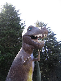
In Oregon, we stopped at a weird place called "Prehistoric Gardens" - basically, a bunch of outdated extinct reptile sculptures in the forest. Well, let's just say that it was only worth our money because of how funky some of the models looked and how bad/off the wall/totally insane some of the information was.
The tyrant king himself welcomes weary drivers in from the road.

The second weirdest depiction of Triceratops I've ever seen.

Pretty standard Ankylosaurus model.

Oh, no! A killer Elasmosaurus (w/ lens flare!) in the middle of the... forest?

This one really screwed with me. OK, I've heard this (obviously) for Oviraptor, but ornithomimids?

My brave companion facing the evil Pteranodon.

Cold blooded? Cretaceaus?
I don't want to know how an Ichthyosaurus made its way into the forest.
In Newport, Oregon, we found the trashiest tourist souvenir shops I've ever seen, which says a lot because I've been to Disneyland, Pier 39, and Fisherman's Wharf. What's worse, these were built right next to the Oregon Coast Aquarium, and named "Aquarium Square Shops" - completely misleading. Anyway, this was literally the first thing we saw when we made it to Newport - a big blue pliosaur.

They didn't even try with this one. They call it "Nessie", the "Yaquina Bay Sea Monster", and "Kronosaurus" all within 4 feet. What?! First off, Nessie is the Loch Ness Monster. Second, I've never heard of a Yaquina Bay Sea Monster (wouldn't be a bay monster?), and it is obviously a sick marketing ploy just like Tahoe Tessie (rhymes with nessie, but at least they bothered to put a 'T' on the beginning). They didn't even bother coming up with an original name (seriously, it doesn't take more than half a second to think of a name other than one used by an already established fake sea monster), AND they go ahead and call it Kronosaurus at the same time.